Copyright
©The Author(s) 2022.
World J Clin Cases. Oct 26, 2022; 10(30): 11066-11073
Published online Oct 26, 2022. doi: 10.12998/wjcc.v10.i30.11066
Published online Oct 26, 2022. doi: 10.12998/wjcc.v10.i30.11066
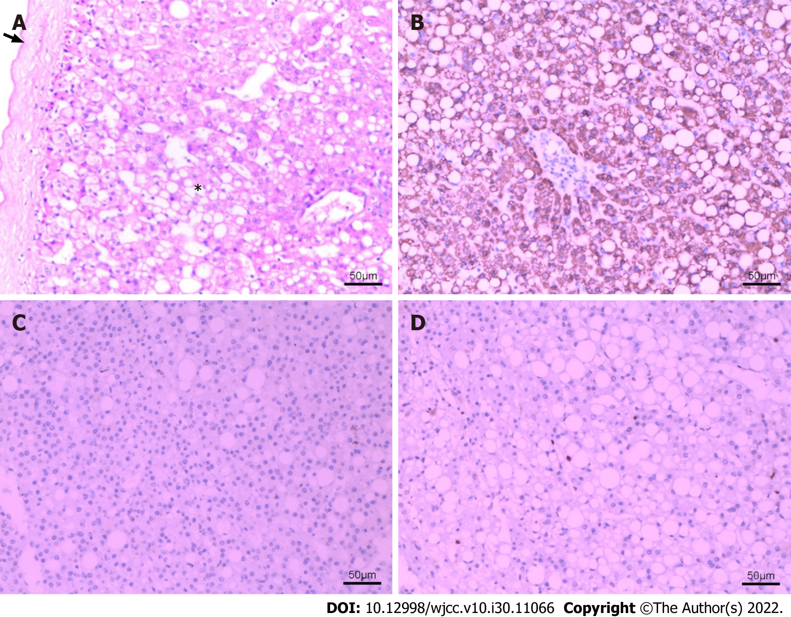
Figure 4 Pathological changes of hepatic steatosis with mass effect.
The HE (hematoxylin and eosin) and immunohistochemical staining of resected specimen at × 100 magnification. A: HE staining of the specimen showed that some normal structures of liver tissue disappeared. No obvious lobular structure was found. There were extensive steatosis of hepatocytes (*), and fibrous cell layer around the lesions (arrow). B: Immunohistochemical staining: Hep Par 1 (hepatocyte paraffin 1) (+); C: Immunohistochemical staining: GPC-3 (glypican-3) (-); D: Immunohistochemical staining: Ki-67 index (2%).
- Citation: Hu N, Su SJ, Li JY, Zhao H, Liu SF, Wang LS, Gong RZ, Li CT. Hepatic steatosis with mass effect: A case report. World J Clin Cases 2022; 10(30): 11066-11073
- URL: https://www.wjgnet.com/2307-8960/full/v10/i30/11066.htm
- DOI: https://dx.doi.org/10.12998/wjcc.v10.i30.11066


