Copyright
©The Author(s) 2022.
World J Clin Cases. Oct 26, 2022; 10(30): 11066-11073
Published online Oct 26, 2022. doi: 10.12998/wjcc.v10.i30.11066
Published online Oct 26, 2022. doi: 10.12998/wjcc.v10.i30.11066
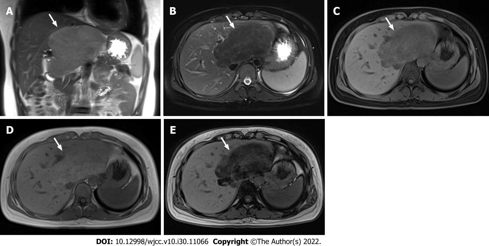
Figure 3 Magnetic resonance imaging of hepatic steatosis with mass effect.
A: Coronal T2-weighted non-fat-saturated image revealed that the lesion (arrow) in the caudate lobe of the liver showed slight hyperintensity and locally protruding beyond the liver outline; B: The signal intensity was reduced on axial T2-weighted fat-saturated images; C: The signal intensity was also decreased on axial T1-weighted fat-saturated images; D: Axial T1-weighted in-phase image showed slight hyperintensity; E: The signal intensity of out-of-phase image decreased significantly. The degree of reduction of the T1-weighted out-of-phase image was higher than that of T1-weighted fat-saturated image.
- Citation: Hu N, Su SJ, Li JY, Zhao H, Liu SF, Wang LS, Gong RZ, Li CT. Hepatic steatosis with mass effect: A case report. World J Clin Cases 2022; 10(30): 11066-11073
- URL: https://www.wjgnet.com/2307-8960/full/v10/i30/11066.htm
- DOI: https://dx.doi.org/10.12998/wjcc.v10.i30.11066


