Copyright
©The Author(s) 2022.
World J Clin Cases. Jan 21, 2022; 10(3): 985-991
Published online Jan 21, 2022. doi: 10.12998/wjcc.v10.i3.985
Published online Jan 21, 2022. doi: 10.12998/wjcc.v10.i3.985
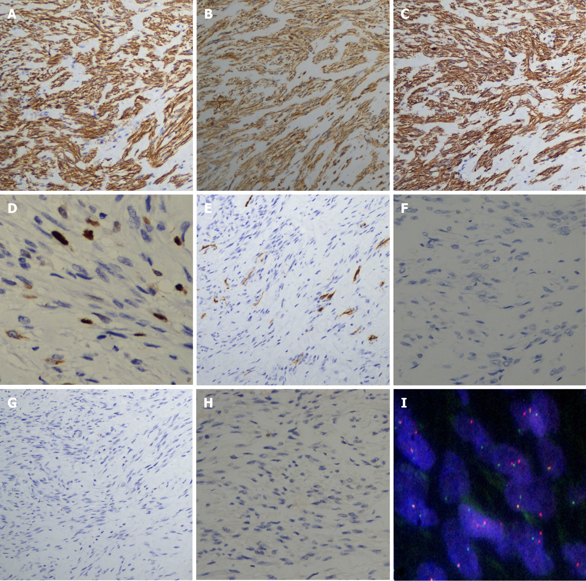
Figure 3 Immunohistochemistry and USP6 gene rearrangement.
A: The spindle cells were positive for smooth muscle actin; B: The spindle cells were positive for vimentin; C: The spindle cells were positive for caldesmon; D: Ki-67 staining of spindle cells revealed a low proliferative index (5%-10%); E: The spindle cells were negative for CD34; F: The spindle cells were negative for S-100; G: The spindle cells were negative for desmin; H: The spindle cells were negative for c-kit; I: Fluorescence in situ hybridization with a separate probe for USP6 indicated that there might be one yellow or red-green adjacent fusion signal and two red-green separation signals in most cells.
- Citation: Meng XH, Liu YC, Xie LS, Huang CP, Xie XP, Fang X. Intravascular fasciitis involving the external jugular vein and subclavian vein: A case report . World J Clin Cases 2022; 10(3): 985-991
- URL: https://www.wjgnet.com/2307-8960/full/v10/i3/985.htm
- DOI: https://dx.doi.org/10.12998/wjcc.v10.i3.985


