Copyright
©The Author(s) 2022.
World J Clin Cases. Jan 21, 2022; 10(3): 856-869
Published online Jan 21, 2022. doi: 10.12998/wjcc.v10.i3.856
Published online Jan 21, 2022. doi: 10.12998/wjcc.v10.i3.856
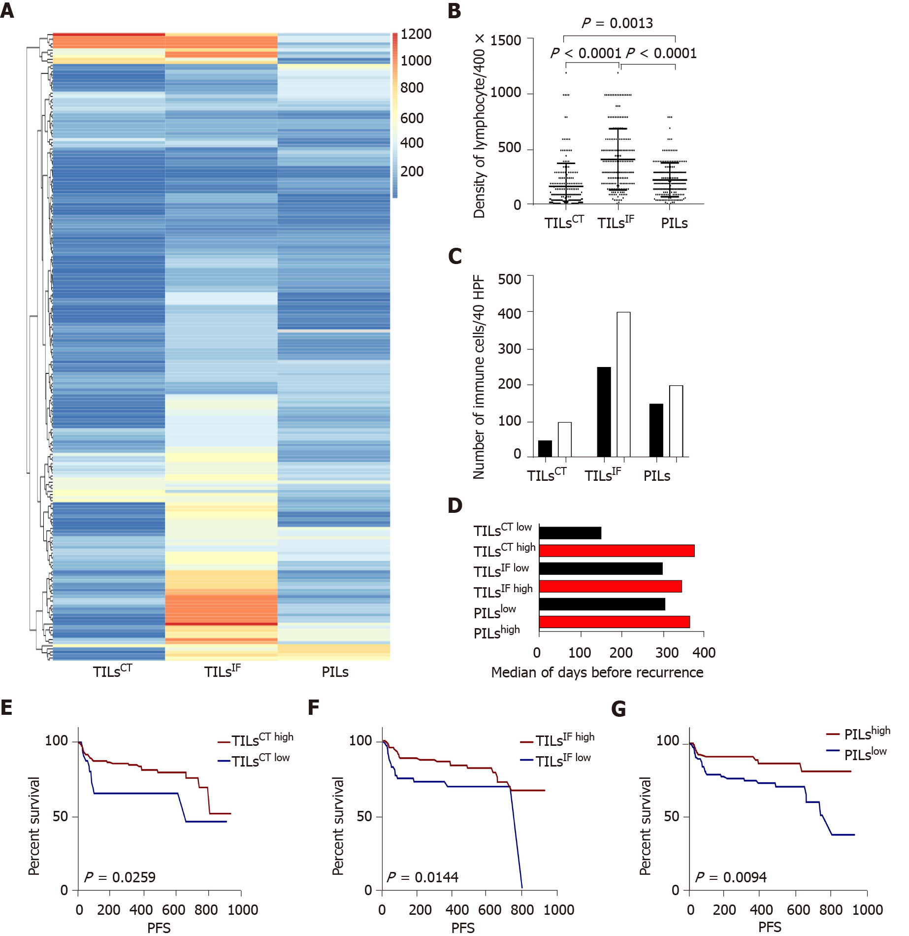
Figure 1 The distribution and recurrence association of tumor infiltrating lymphocytes in the tumor center, invasive front, and peritumor.
A: The spectrum of tumor infiltrating lymphocytes in the tumor center (TILsCT), TILs in the invasive front (TILsIF), and TILs in the peritumor (PILs) of 204 cases. TILsCT were lower than TILsIF and PILs. TILsIF was the most flaming part than the other two areas; B: Dot map of TILsCT, TILsIF, and PILs, indicating a heterogeneous distribution of inflammation; C: Comparison of the mean of TILsCT, TILsIF and PILs from patients with tumor recurrence (black bars) or without tumor recurrence (white bars); D: Median survival time for all patients, with high densities (red bars) or low densities (black bars) of TILsCT, TILsIF and PILs; E, F and G: Patients with high TILsCT, TILsIF, and PILs had a lower recurrence rate. TILsCT: Tumor infiltrating lymphocytes in the tumor center; TILsIF: Tumor infiltrating lymphocytes in the invasive front 1 mm spacing from the malignant nests; PILs: infiltrating lymphocytes in the peritumor.
- Citation: Du M, Cai YM, Yin YL, Xiao L, Ji Y. Evaluating tumor-infiltrating lymphocytes in hepatocellular carcinoma using hematoxylin and eosin-stained tumor sections. World J Clin Cases 2022; 10(3): 856-869
- URL: https://www.wjgnet.com/2307-8960/full/v10/i3/856.htm
- DOI: https://dx.doi.org/10.12998/wjcc.v10.i3.856


