Copyright
©The Author(s) 2022.
World J Clin Cases. Jan 21, 2022; 10(3): 1099-1105
Published online Jan 21, 2022. doi: 10.12998/wjcc.v10.i3.1099
Published online Jan 21, 2022. doi: 10.12998/wjcc.v10.i3.1099
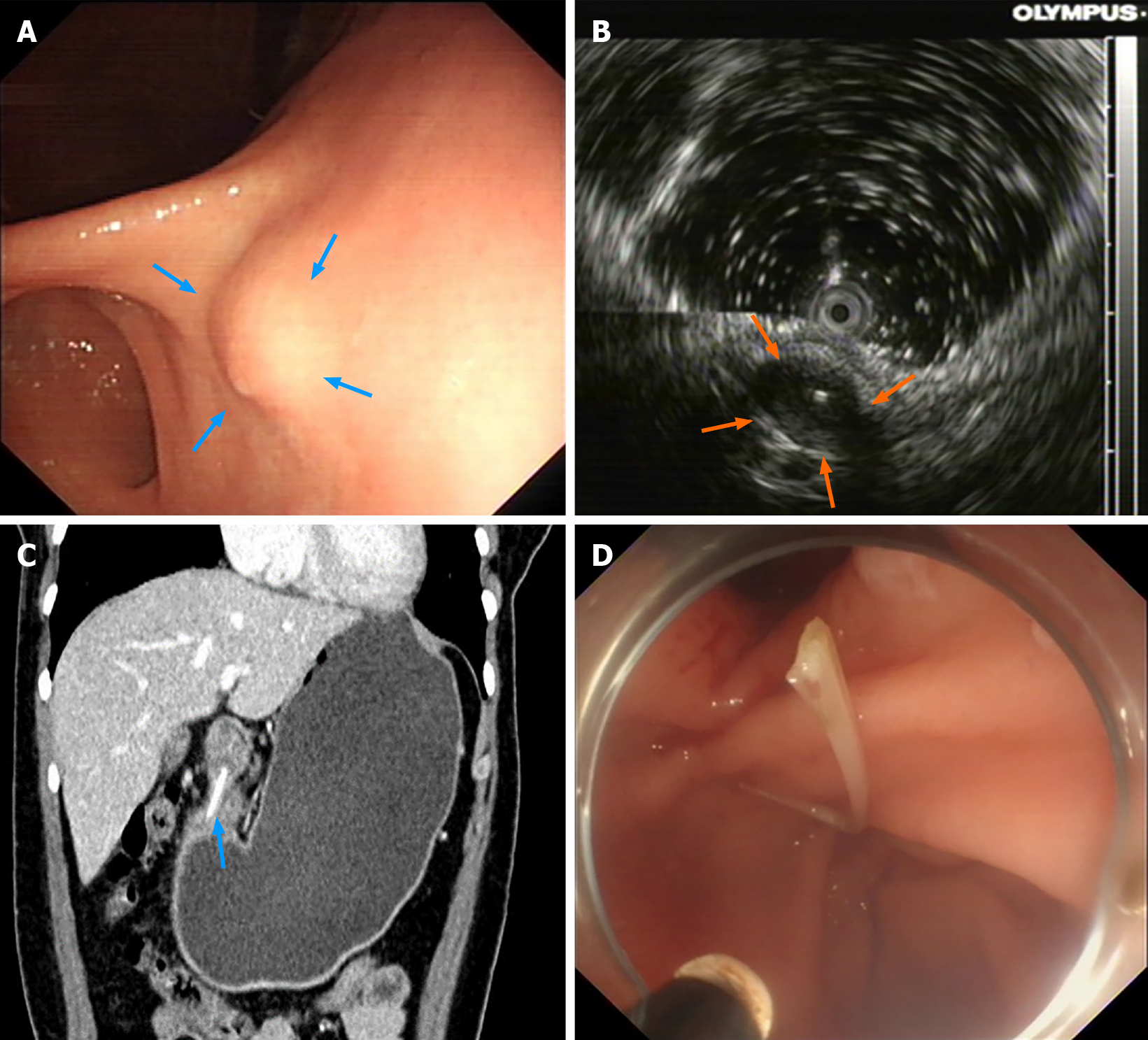
Figure 1 Findings from endoscopy and a computed tomography scan during the diagnostic process and endoscopic treatment.
A: Endoscopy revealed an elevated lesion in the gastric antrum (blue arrow); B: Endoscopic ultrasonography showing a hypoechoic mass in the posterior wall of the gastric antrum (orange arrow); C: Abdominal computed tomography (CT) scan showing a hyperdense linear structure in the gastric antrum wall (blue arrow), CT value: 968 HU; D: During endoscopic surgery, an L-shape fish bone was removed from the lesion.
- Citation: Li J, Wang QQ, Xue S, Zhang YY, Xu QY, Zhang XH, Feng L. Gastric submucosal lesion caused by an embedded fish bone: A case report. World J Clin Cases 2022; 10(3): 1099-1105
- URL: https://www.wjgnet.com/2307-8960/full/v10/i3/1099.htm
- DOI: https://dx.doi.org/10.12998/wjcc.v10.i3.1099


