Copyright
©The Author(s) 2022.
World J Clin Cases. Jan 21, 2022; 10(3): 1041-1049
Published online Jan 21, 2022. doi: 10.12998/wjcc.v10.i3.1041
Published online Jan 21, 2022. doi: 10.12998/wjcc.v10.i3.1041
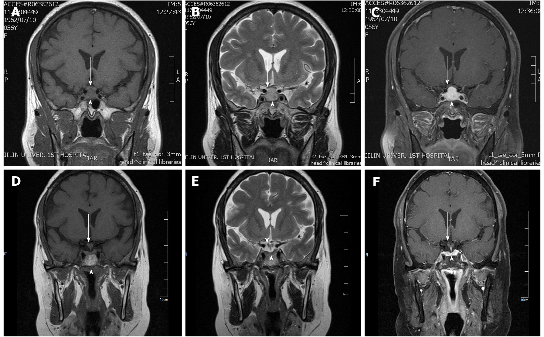
Figure 1 Pretreatment coronal magnetic resonance imaging showing pituitary enlargement (arrowhead) and optic chiasm elevation (arrow).
A: In the T1 sequence; B: In the T2 sequence; C: Postgadolinium-enhanced coronal magnetic resonance imaging showing an enlarged pituitary gland with significant homogeneous enhancement (arrowhead) and an elevation of the optic chiasm (arrow); D: Posttreatment coronal magnetic resonance imaging showing an almost normal pituitary gland in the T1 sequence; E: Posttreatment coronal magnetic resonance imaging showing an almost normal pituitary gland in the T2 sequence; F: With gadolinium enhancement in the coronal position.
- Citation: Yang MG, Cai HQ, Wang SS, Liu L, Wang CM. Full recovery from chronic headache and hypopituitarism caused by lymphocytic hypophysitis: A case report. World J Clin Cases 2022; 10(3): 1041-1049
- URL: https://www.wjgnet.com/2307-8960/full/v10/i3/1041.htm
- DOI: https://dx.doi.org/10.12998/wjcc.v10.i3.1041


