Copyright
©The Author(s) 2022.
World J Clin Cases. Jan 21, 2022; 10(3): 1032-1040
Published online Jan 21, 2022. doi: 10.12998/wjcc.v10.i3.1032
Published online Jan 21, 2022. doi: 10.12998/wjcc.v10.i3.1032
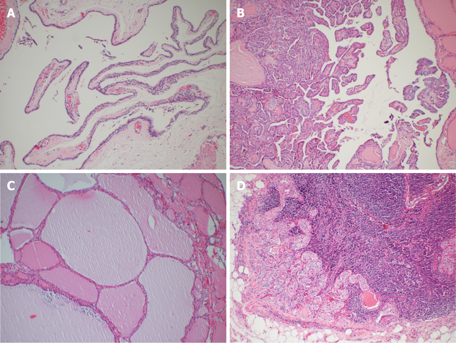
Figure 4 Histopathological examinations.
A: Parathyroid cyst: a single locular cystic mass covered by a single layer of flattened transparent cells with small clusters of extruded parathyroid tissue in the wall; B: Thyroid papillary carcinoma: complex branching papilla with fibrous vascular center, and surface coated with simple columnar epithelium. The epithelial nuclei were ground-glass like, with nuclear grooves, intranuclear pseudo-inclusions, and nuclear overlap; C: Nodular goiter: the follicles vary in size and are filled with colloid; D: Metastatic lesions of thyroid papillary carcinoma in lymph nodes. Hematoxylin and eosin staining, 100× magnification.
- Citation: Xu JL, Dong S, Sun LL, Zhu JX, Liu J. Multiple endocrine neoplasia type 1 combined with thyroid neoplasm: A case report and review of literatures. World J Clin Cases 2022; 10(3): 1032-1040
- URL: https://www.wjgnet.com/2307-8960/full/v10/i3/1032.htm
- DOI: https://dx.doi.org/10.12998/wjcc.v10.i3.1032


