Copyright
©The Author(s) 2022.
World J Clin Cases. Jan 21, 2022; 10(3): 1032-1040
Published online Jan 21, 2022. doi: 10.12998/wjcc.v10.i3.1032
Published online Jan 21, 2022. doi: 10.12998/wjcc.v10.i3.1032
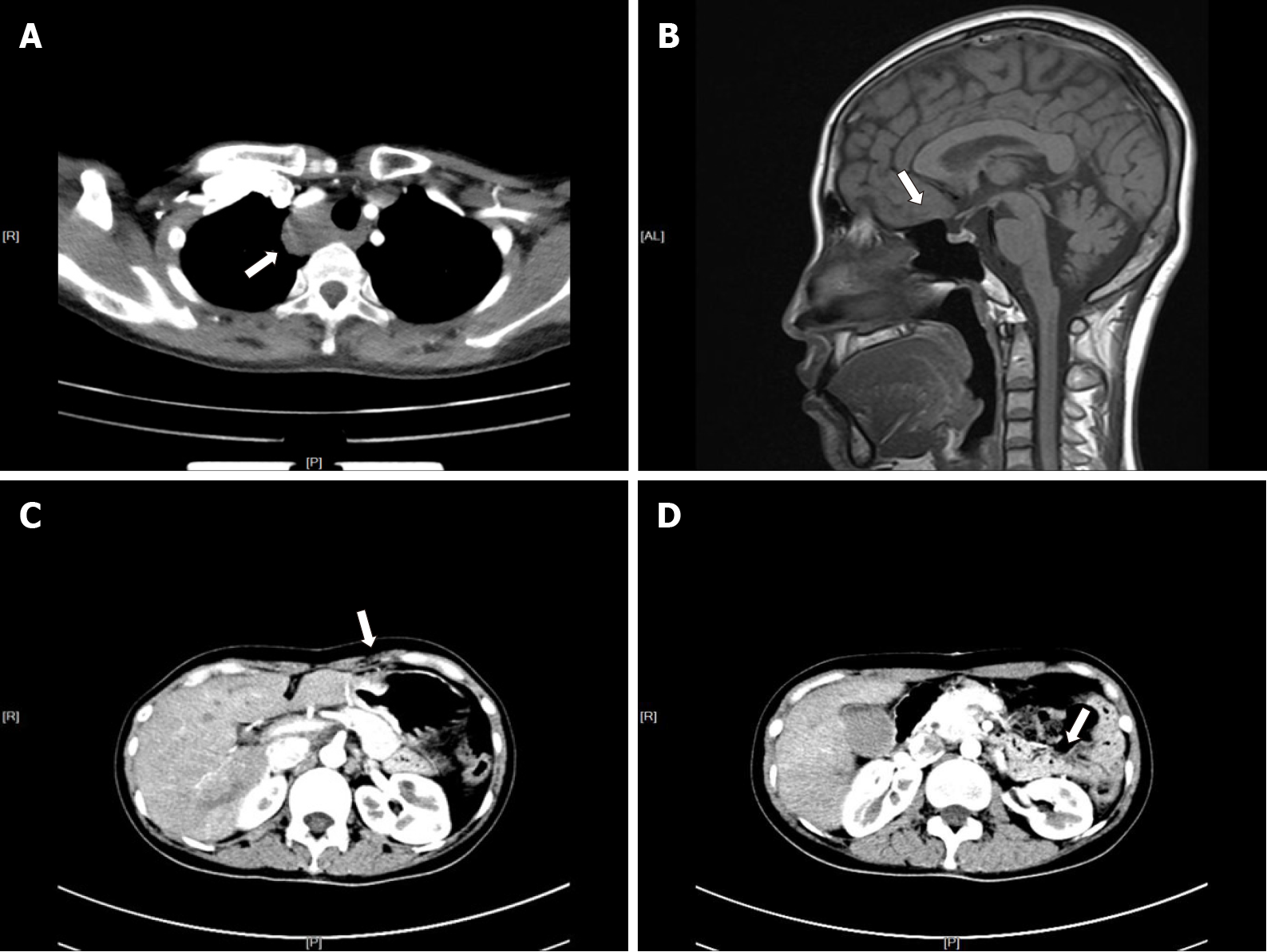
Figure 2 Computed tomography/magnetic resonance imaging examination.
A: A lesion located in the right side of the trachea (white arrow); B: Enlarged pituitary structure (white arrow); C: Remnant stomach anastomosed to the jejunum (white arrow); D: Remnant pancreas body and tail (white arrow).
- Citation: Xu JL, Dong S, Sun LL, Zhu JX, Liu J. Multiple endocrine neoplasia type 1 combined with thyroid neoplasm: A case report and review of literatures. World J Clin Cases 2022; 10(3): 1032-1040
- URL: https://www.wjgnet.com/2307-8960/full/v10/i3/1032.htm
- DOI: https://dx.doi.org/10.12998/wjcc.v10.i3.1032


