Copyright
©The Author(s) 2022.
World J Clin Cases. Jan 21, 2022; 10(3): 1016-1023
Published online Jan 21, 2022. doi: 10.12998/wjcc.v10.i3.1016
Published online Jan 21, 2022. doi: 10.12998/wjcc.v10.i3.1016
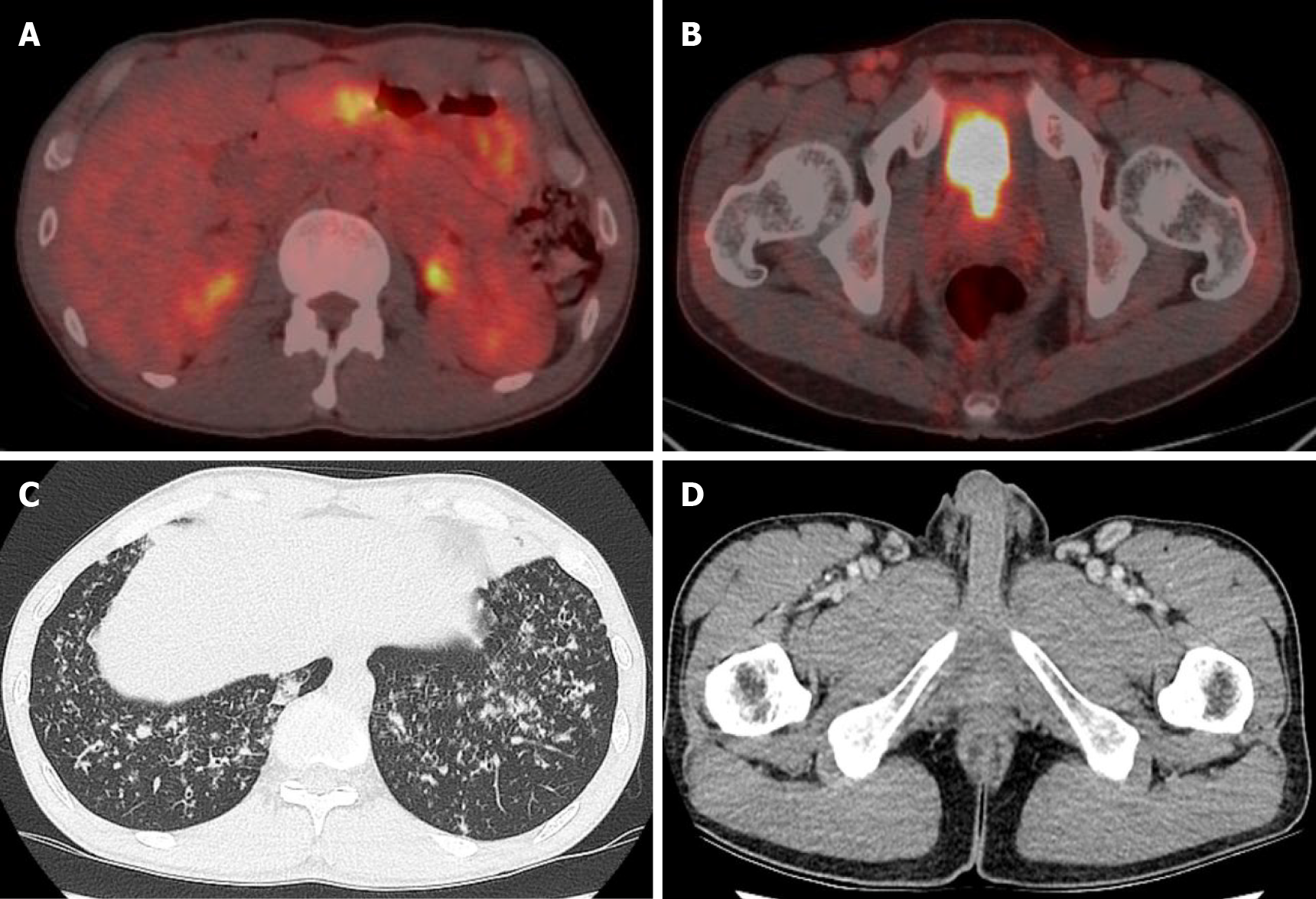
Figure 1 The positron emission tomography scan, chest computed tomography, and abdominal computed tomography.
A and B: Diffuse hypermetabolic activity in the gastric body and mild increased metabolic activity in both inguinal areas found on an axial view of the positron emission tomography scan; C: In the axial view of the chest computed tomography (CT), ground glass opacities and centrilobular nodules were found in both lung fields; D: Abdominal CT showing multiple enlarged lymph nodes in the bilateral inguinal area.
- Citation: Kim J, Kim YS, Lee HJ, Park SG. Pulmonary amyloidosis and multiple myeloma mimicking lymphoma in a patient with Sjogren’s syndrome: A case report. World J Clin Cases 2022; 10(3): 1016-1023
- URL: https://www.wjgnet.com/2307-8960/full/v10/i3/1016.htm
- DOI: https://dx.doi.org/10.12998/wjcc.v10.i3.1016


