Copyright
©The Author(s) 2022.
World J Clin Cases. Jan 21, 2022; 10(3): 1000-1007
Published online Jan 21, 2022. doi: 10.12998/wjcc.v10.i3.1000
Published online Jan 21, 2022. doi: 10.12998/wjcc.v10.i3.1000
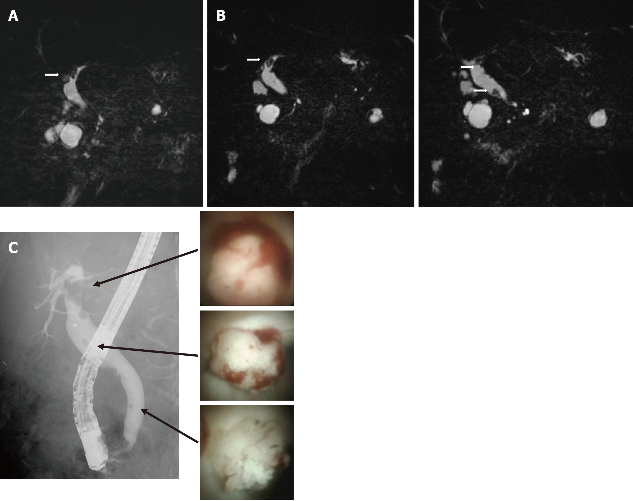
Figure 3 Follow-up imaging after diagnosis.
A: Six months after magnetic resonance cholangiopancreatography (MRCP) showing a defect of the left intrahepatic duct. B: One year after MRCP showing a defect in the left intrahepatic duct and multiple defects in the extrahepatic duct. C: Endoscopic retrograde cholangiography showing multiple filling defects of contrast agent in the extrahepatic and intrahepatic ducts. Peroral cholangioscopy showing papillary tumors in the intrahepatic and extrahepatic ducts.
- Citation: Fukuya H, Kuwano A, Nagasawa S, Morita Y, Tanaka K, Yada M, Masumoto A, Motomura K. Multicentric recurrence of intraductal papillary neoplasm of bile duct after spontaneous detachment of primary tumor: A case report. World J Clin Cases 2022; 10(3): 1000-1007
- URL: https://www.wjgnet.com/2307-8960/full/v10/i3/1000.htm
- DOI: https://dx.doi.org/10.12998/wjcc.v10.i3.1000


