Copyright
©The Author(s) 2022.
World J Clin Cases. Jan 21, 2022; 10(3): 1000-1007
Published online Jan 21, 2022. doi: 10.12998/wjcc.v10.i3.1000
Published online Jan 21, 2022. doi: 10.12998/wjcc.v10.i3.1000
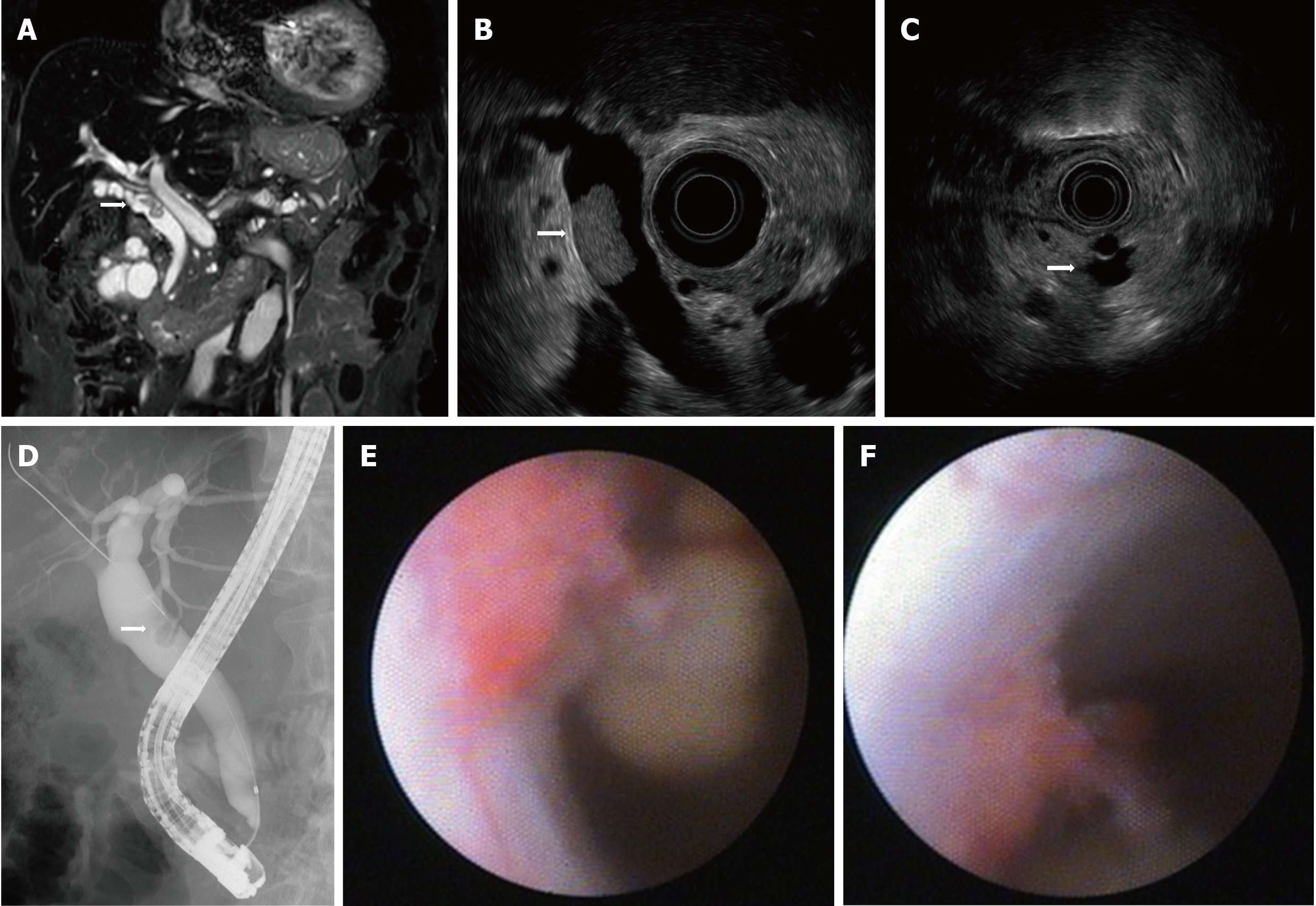
Figure 1 Imaging results upon referral.
A: Magnetic resonance cholangiopancreatography showing a filling defect in the common bile duct (CBD); B: Endoscopic ultrasound (EUS) showing a papillary tumor in the CBD; C: EUS showing intraductal papillary mucinous neoplasm with a mural nodule; D: ERC showing a filling defect of contrast agent in the CBD; E: Peroral cholangioscopy showing a papillary tumor in the CBD; F: Tumor spontaneously detached during examination.
- Citation: Fukuya H, Kuwano A, Nagasawa S, Morita Y, Tanaka K, Yada M, Masumoto A, Motomura K. Multicentric recurrence of intraductal papillary neoplasm of bile duct after spontaneous detachment of primary tumor: A case report. World J Clin Cases 2022; 10(3): 1000-1007
- URL: https://www.wjgnet.com/2307-8960/full/v10/i3/1000.htm
- DOI: https://dx.doi.org/10.12998/wjcc.v10.i3.1000


