Copyright
©The Author(s) 2022.
World J Clin Cases. Oct 16, 2022; 10(29): 10803-10810
Published online Oct 16, 2022. doi: 10.12998/wjcc.v10.i29.10803
Published online Oct 16, 2022. doi: 10.12998/wjcc.v10.i29.10803
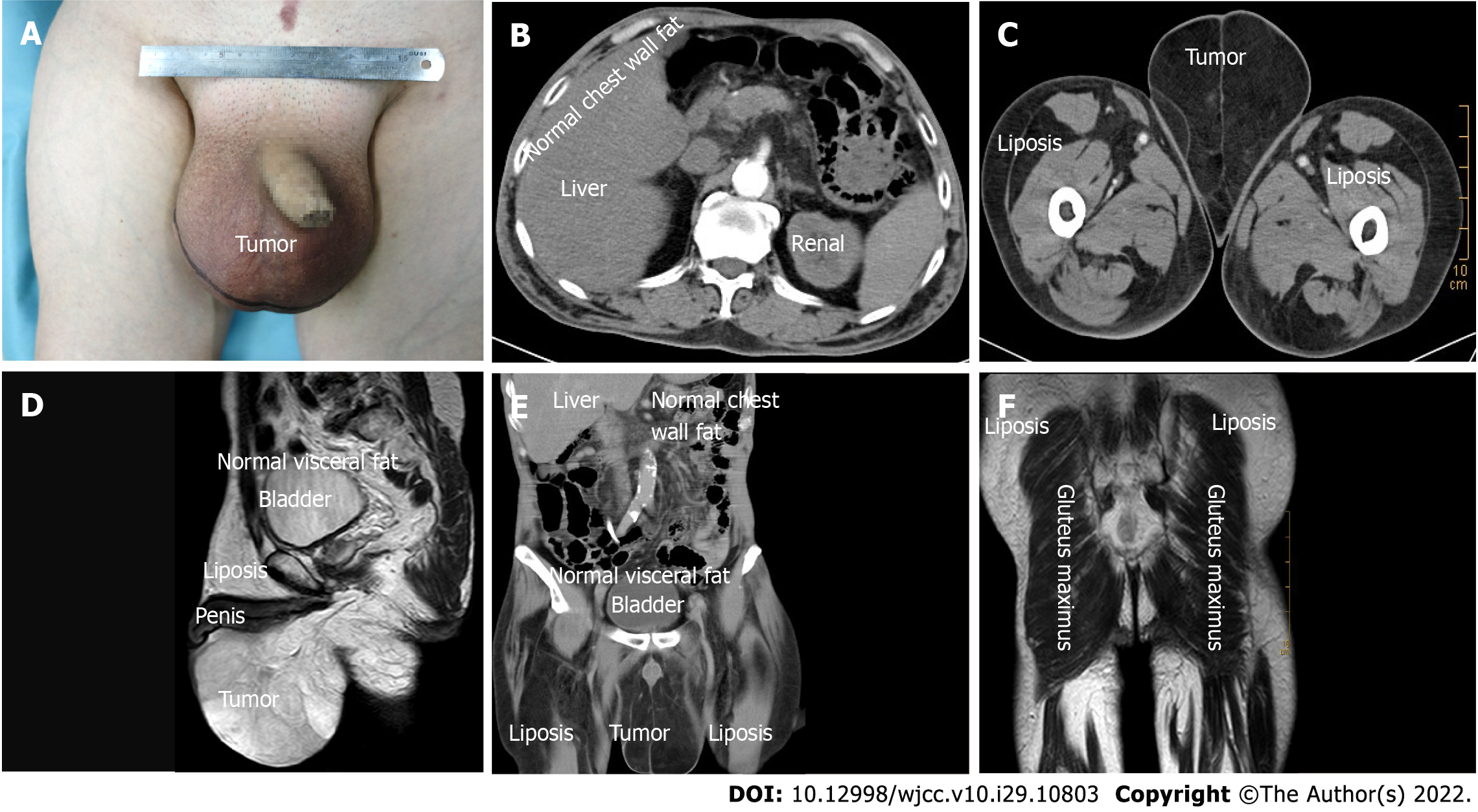
Figure 1 Bilateral scrotal lipoma imaging examination findings.
A: Preoperative scrotal lipoma image; B-D: The computer tomography images showed liposis of the lower abdomen, perineum and thigh but no liposis in the chest wall or pelvic cavity; E and F: Magnetic resonance imaging image showing the same findings.
- Citation: Chen Y, Li XN, Yi XL, Tang Y. Giant bilateral scrotal lipoma with abnormal somatic fat distribution: A case report. World J Clin Cases 2022; 10(29): 10803-10810
- URL: https://www.wjgnet.com/2307-8960/full/v10/i29/10803.htm
- DOI: https://dx.doi.org/10.12998/wjcc.v10.i29.10803


