Copyright
©The Author(s) 2022.
World J Clin Cases. Oct 16, 2022; 10(29): 10787-10793
Published online Oct 16, 2022. doi: 10.12998/wjcc.v10.i29.10787
Published online Oct 16, 2022. doi: 10.12998/wjcc.v10.i29.10787
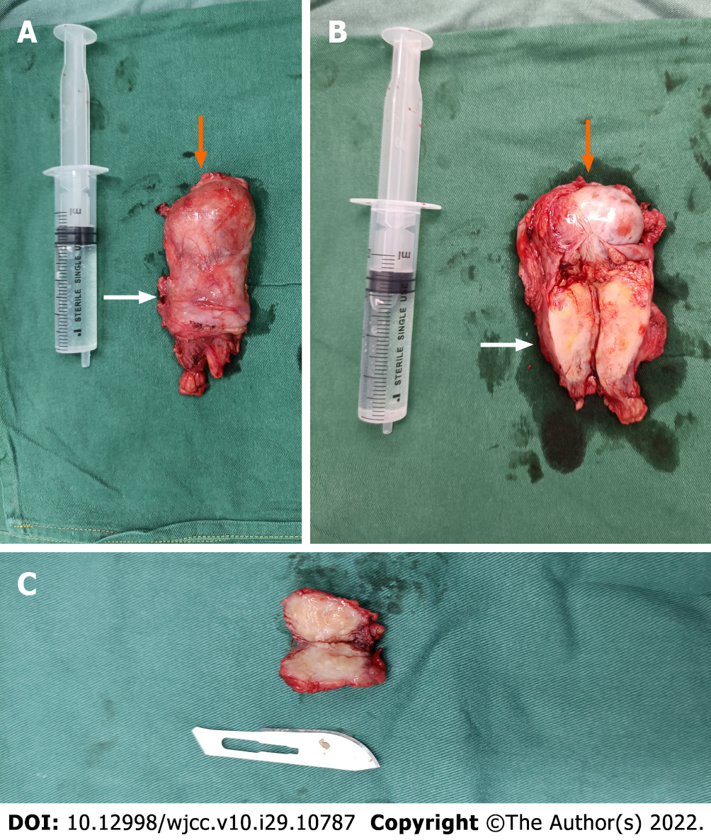
Figure 3 Photograph of the scrotal mass.
A: The left testis (orange arrow); B: The left spermatic cord mass (white arrow); C: The mass in the tail of the right epididymis. The size of the syringe is 20 mL.
- Citation: Lv DY, Xie HJ, Cui F, Zhou HY, Shuang WB. Bilateral occurrence of sperm granulomas in the left spermatic cord and on the right epididymis: A case report. World J Clin Cases 2022; 10(29): 10787-10793
- URL: https://www.wjgnet.com/2307-8960/full/v10/i29/10787.htm
- DOI: https://dx.doi.org/10.12998/wjcc.v10.i29.10787


