Copyright
©The Author(s) 2022.
World J Clin Cases. Oct 16, 2022; 10(29): 10787-10793
Published online Oct 16, 2022. doi: 10.12998/wjcc.v10.i29.10787
Published online Oct 16, 2022. doi: 10.12998/wjcc.v10.i29.10787
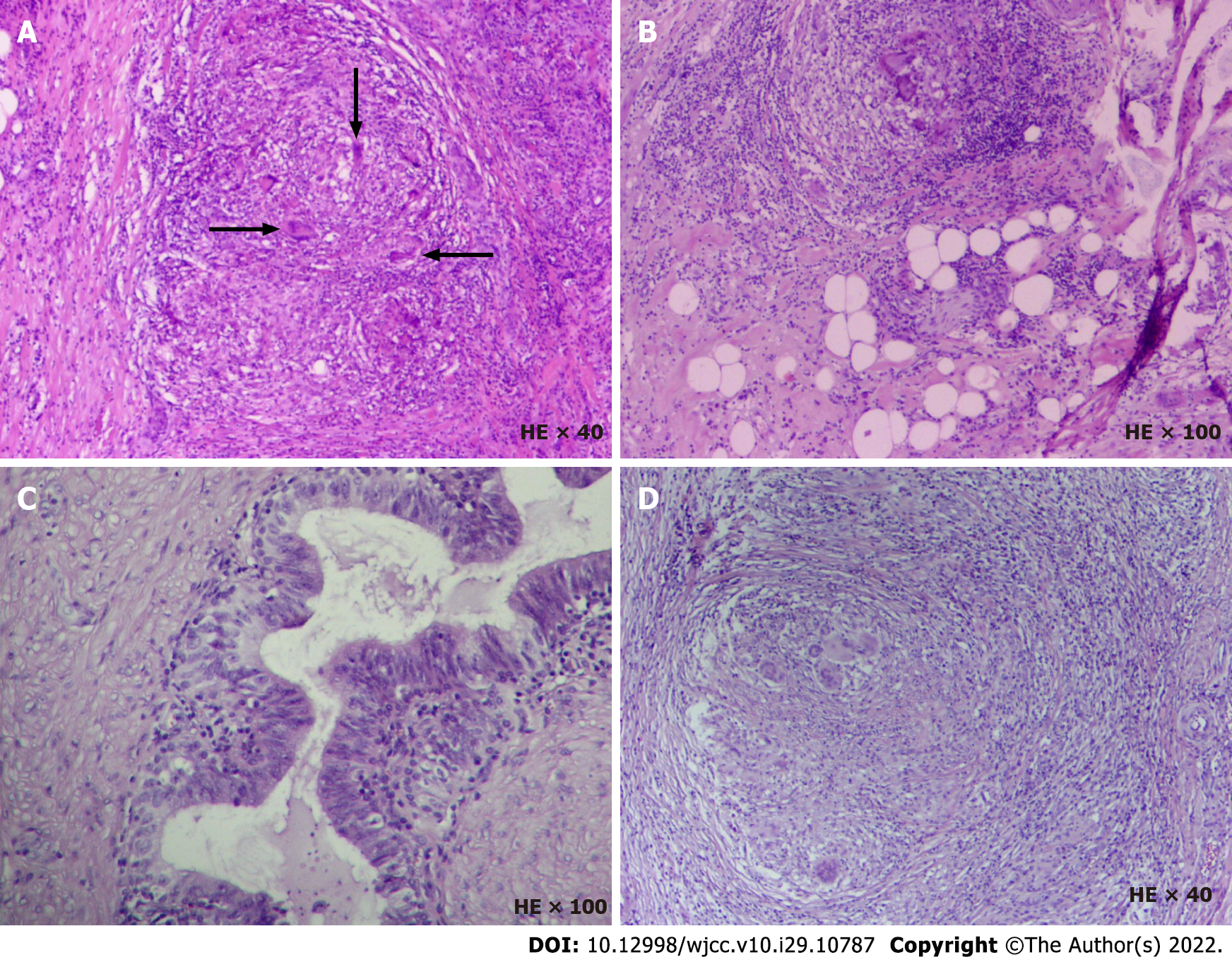
Figure 1 Histopathological analysis of the resected specimen.
A and B: The left scrotum. Spermatic cord mass is consistent with granulomatous lesion with little necrosis. Macrophages can be seen to engulf degraded sperm (arrow), considered as sperm granuloma. The incisal margin did not change significantly. There was no significant change in testis and epididymis; C and D: The right epididymis was infiltrated with lymphocytes, mononuclear cells and eosinophils. The formation of an epithelioid granuloma was found in the epididymis along with multinucleated giant cells, including foreign body giant cells and Langhans giant cells. Coagulative necrosis was found in some areas along with complete necrosis of small focus which is consistent with granulomatous lesions. HE: Hematoxylin-eosin staining.
- Citation: Lv DY, Xie HJ, Cui F, Zhou HY, Shuang WB. Bilateral occurrence of sperm granulomas in the left spermatic cord and on the right epididymis: A case report. World J Clin Cases 2022; 10(29): 10787-10793
- URL: https://www.wjgnet.com/2307-8960/full/v10/i29/10787.htm
- DOI: https://dx.doi.org/10.12998/wjcc.v10.i29.10787


