Copyright
©The Author(s) 2022.
World J Clin Cases. Oct 16, 2022; 10(29): 10772-10778
Published online Oct 16, 2022. doi: 10.12998/wjcc.v10.i29.10772
Published online Oct 16, 2022. doi: 10.12998/wjcc.v10.i29.10772
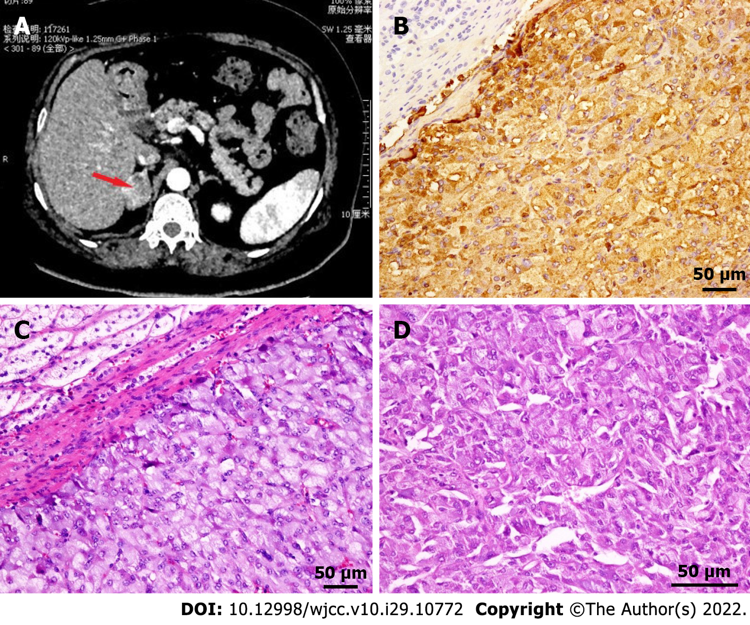
Figure 3 Adrenal computed tomography scan and pathology.
A: Mass-like soft tissue shadow with heterogeneous density was present in the medial branch of the right adrenal gland; B: Immunohistochemical staining showed Chromogranin A (+), which was confined to the adrenal gland and did not invade the peri-adrenal tissue; C: HE stained tissue observed under 200 × light microscope; D: HE stained tissue observed under 400 × light microscope. HE: Hematoxylin-eosin.
- Citation: Wang ZH, Fan JR, Zhang GY, Li XL, Li L. Atypical Takotsubo cardiomyopathy presenting as acute coronary syndrome: A case report. World J Clin Cases 2022; 10(29): 10772-10778
- URL: https://www.wjgnet.com/2307-8960/full/v10/i29/10772.htm
- DOI: https://dx.doi.org/10.12998/wjcc.v10.i29.10772


