Copyright
©The Author(s) 2022.
World J Clin Cases. Oct 16, 2022; 10(29): 10763-10771
Published online Oct 16, 2022. doi: 10.12998/wjcc.v10.i29.10763
Published online Oct 16, 2022. doi: 10.12998/wjcc.v10.i29.10763
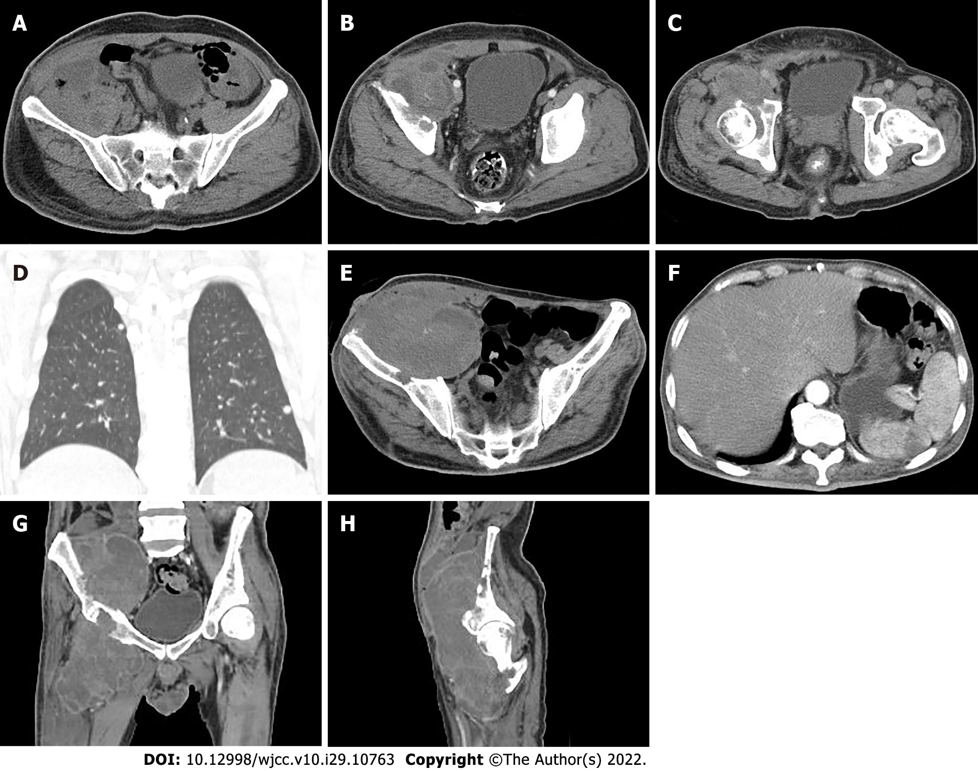
Figure 6 Computed tomography monitoring after chemotherapy.
A-D: 1 mo and 7 d after the initial diagnosis; A: The lesion was larger in size than before and the adjacent abdominal wall was unevenly thickened; B: There were an increased amounts of necrosis lesions; C: The lesion invaded the acetabulum; D: Small and round solid nodules were found in both lung lobes; E-F: 5 mo and 20 d after the initial diagnosis; E: Thinning of the right abdominal wall with a skin fistula was caused by the mass; F: A patchy, hypodense infarcted area in the spleen represented a metastatic lesion; G: Further enlargement of the lesion; and H: Multiple osteolytic destruction was shown in the right ilium, acetabulum, and proximal femur.
- Citation: Huang WP, Gao G, Yang Q, Chen Z, Qiu YK, Gao JB, Kang L. Malignant giant cell tumors of the tendon sheath of the right hip: A case report. World J Clin Cases 2022; 10(29): 10763-10771
- URL: https://www.wjgnet.com/2307-8960/full/v10/i29/10763.htm
- DOI: https://dx.doi.org/10.12998/wjcc.v10.i29.10763


