Copyright
©The Author(s) 2022.
World J Clin Cases. Oct 16, 2022; 10(29): 10763-10771
Published online Oct 16, 2022. doi: 10.12998/wjcc.v10.i29.10763
Published online Oct 16, 2022. doi: 10.12998/wjcc.v10.i29.10763
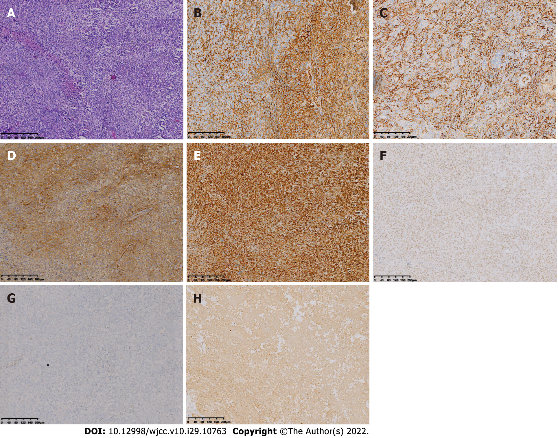
Figure 5 Histopathological images.
A: Hematoxylin-eosin (HE) staining showed that the tumor was composed of mononuclear synovial-like cells and sarcoma cells. Lacunar-like lacunae were shown within the tumor cells and pleomorphism and heterogeneity of the tumor cells were obvious. Pathological nuclear division was seen and a collagen matrix was found between the cells (magnification ×100); B: Immunohistochemical staining revealed CD31 positivity (Envision, 100×); C: Immunohistochemical staining revealed CD34 positivity (Envision, 100×); D: Immunohistochemical staining revealed CD99 positivity (Envision, 100×); E: Immunohistochemical staining revealed Vimentin positivity (Envision, 100×); F: Immunohistochemical staining revealed INI-1 positivity (Envision, 100×); G: Immunohistochemical staining revealed TLE1 positivity (Envision, 100×); H: Immunohistochemical staining revealed CK positivity (Envision, 100×).
- Citation: Huang WP, Gao G, Yang Q, Chen Z, Qiu YK, Gao JB, Kang L. Malignant giant cell tumors of the tendon sheath of the right hip: A case report. World J Clin Cases 2022; 10(29): 10763-10771
- URL: https://www.wjgnet.com/2307-8960/full/v10/i29/10763.htm
- DOI: https://dx.doi.org/10.12998/wjcc.v10.i29.10763


