Copyright
©The Author(s) 2022.
World J Clin Cases. Oct 16, 2022; 10(29): 10708-10712
Published online Oct 16, 2022. doi: 10.12998/wjcc.v10.i29.10708
Published online Oct 16, 2022. doi: 10.12998/wjcc.v10.i29.10708
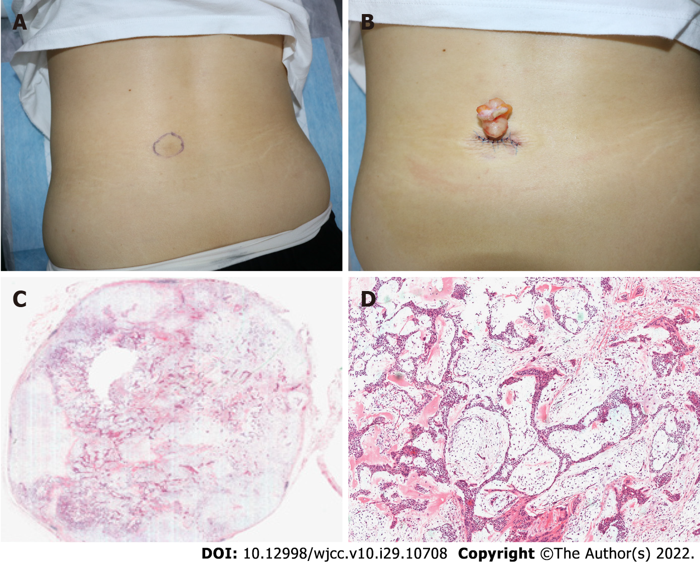
Figure 1 Images of the mass.
A: A Clinical image of a subcutaneous mass, ranging from 3-4 cm in diameter; B: A yellow, smooth, tough mass with a clear boundary and a size of about 5 cm × 4 cm; C: A well-defined dermal tumor (hematoxylin-eosin staining, × 10); D: Tumor with nests, sheets, and cords of basal-like cells, glandular structures, interstitial mucin deposition, and chondroid structures in some areas (hematoxylin-eosin staining, × 200).
- Citation: Huang QF, Shao Y, Yu B, Hu XP. Chondroid syringoma of the lower back simulating lipoma: A case report. World J Clin Cases 2022; 10(29): 10708-10712
- URL: https://www.wjgnet.com/2307-8960/full/v10/i29/10708.htm
- DOI: https://dx.doi.org/10.12998/wjcc.v10.i29.10708


