Copyright
©The Author(s) 2022.
World J Clin Cases. Oct 16, 2022; 10(29): 10663-10669
Published online Oct 16, 2022. doi: 10.12998/wjcc.v10.i29.10663
Published online Oct 16, 2022. doi: 10.12998/wjcc.v10.i29.10663
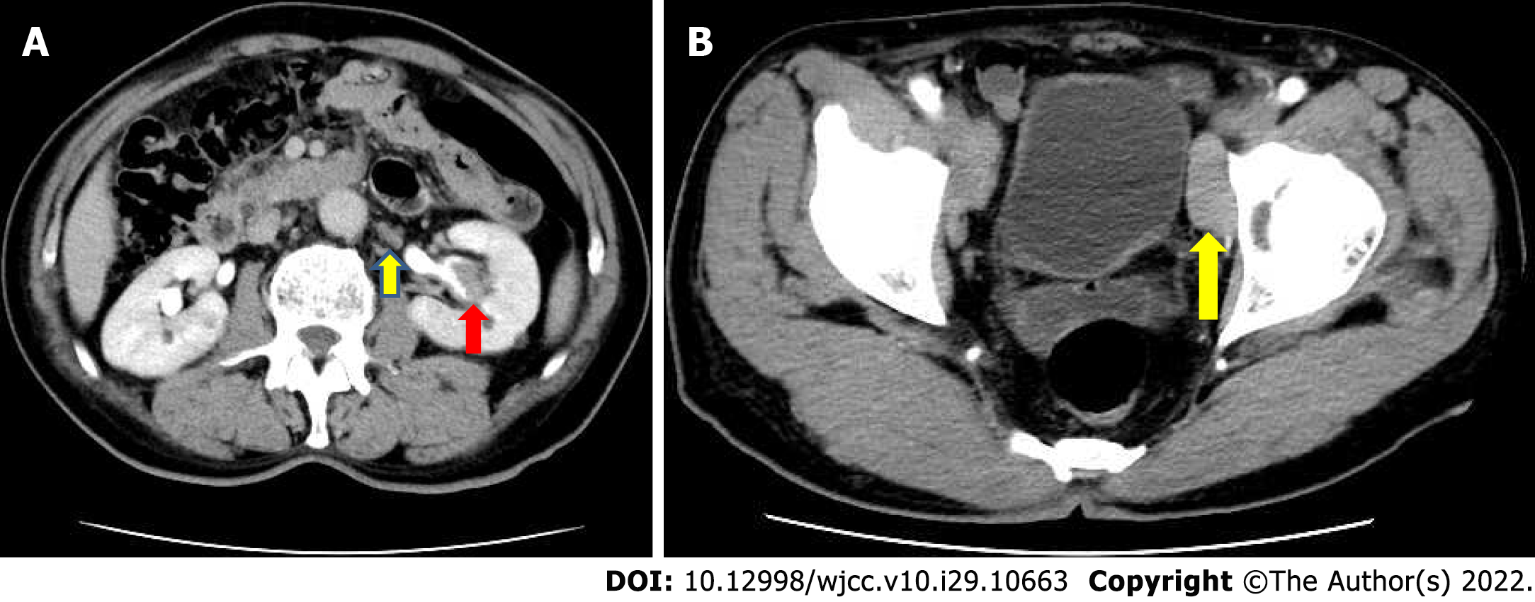
Figure 1 Multiple enlarged lymph nodes were noted in the retroperitoneal and pelvic walls.
A: Abdominal computed tomography imaging demonstrated a tumor mass of approximately 2.7 cm × 1.5 cm × 1 cm in the central part of the left renal pelvis cavity; B: Multiple enlarged lymph nodes were noted in the retroperitoneal and pelvic walls.
- Citation: Yang HJ, Huang X. Synchronous renal pelvis carcinoma associated with small lymphocytic lymphoma: A case report. World J Clin Cases 2022; 10(29): 10663-10669
- URL: https://www.wjgnet.com/2307-8960/full/v10/i29/10663.htm
- DOI: https://dx.doi.org/10.12998/wjcc.v10.i29.10663


