Copyright
©The Author(s) 2022.
World J Clin Cases. Oct 16, 2022; 10(29): 10647-10654
Published online Oct 16, 2022. doi: 10.12998/wjcc.v10.i29.10647
Published online Oct 16, 2022. doi: 10.12998/wjcc.v10.i29.10647
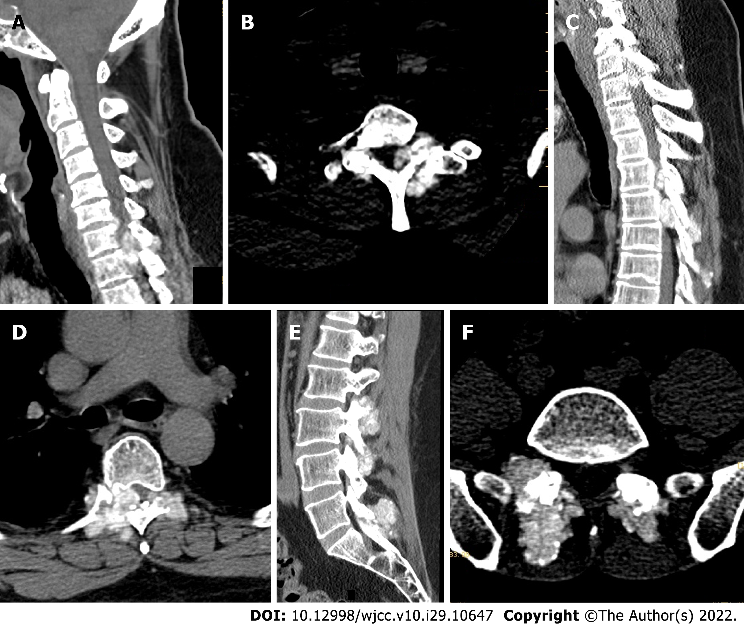
Figure 1 Preoperative computed tomography of the cervical, thoracic, lumbar, and sacral spine.
A-D: Multiple tophi in the whole spine, with spinal cord compression and degeneration at C7/T1, T5, and T6 Levels; E and F: Multiple abnormal high signals that tophi in the lumbar and sacral vertebral subtalar joints, and tophus deposits.
- Citation: Chen HJ, Chen DY, Zhou SZ, Chi KD, Wu JZ, Huang FL. Multiple tophi deposits in the spine: A case report. World J Clin Cases 2022; 10(29): 10647-10654
- URL: https://www.wjgnet.com/2307-8960/full/v10/i29/10647.htm
- DOI: https://dx.doi.org/10.12998/wjcc.v10.i29.10647


