Copyright
©The Author(s) 2022.
World J Clin Cases. Oct 16, 2022; 10(29): 10638-10646
Published online Oct 16, 2022. doi: 10.12998/wjcc.v10.i29.10638
Published online Oct 16, 2022. doi: 10.12998/wjcc.v10.i29.10638
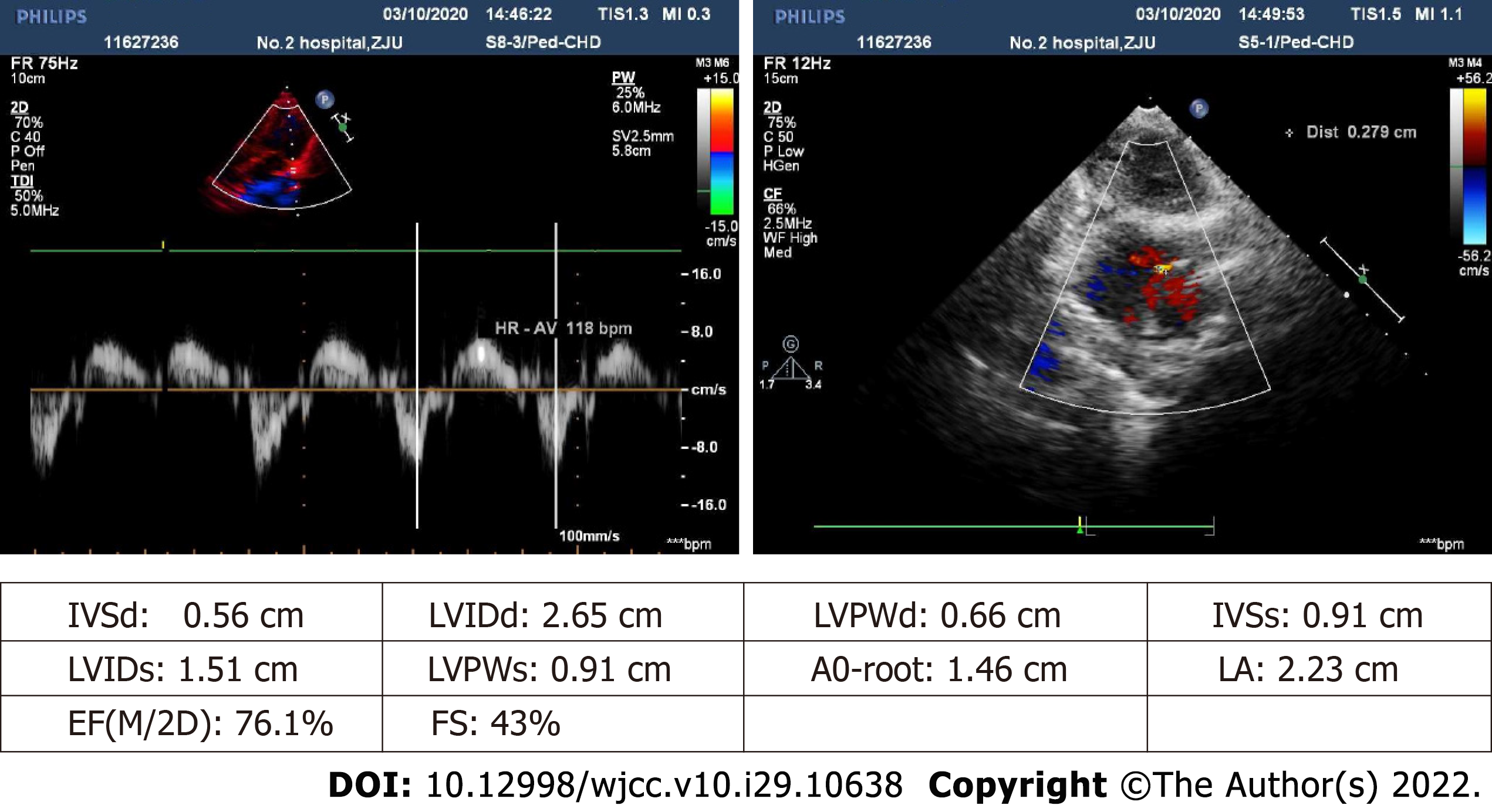
Figure 1 Echocardiographic imaging prior to surgery.
Atrial and ventricular chambers are of normal size (no abnormal internal echoes) and expected thickness, with visible 0.3-cm echoic interruption of middle and lower atrial septum (slightly less than in the prior year). Color Doppler flow imaging (CDFI) reveals left-to-right red-colored streamers, shunt velocity not measured. Interventricular septum remains structurally intact. Valvular echoes appear satisfactory (opening/closing normally), exhibiting trace tricuspid systolic regurgitation (multicolored, mainly blue) by CDFI.
- Citation: Liu L, Chen P, Fang LL, Yu LN. Perioperative anesthesia management in pediatric liver transplant recipient with atrial septal defect: A case report. World J Clin Cases 2022; 10(29): 10638-10646
- URL: https://www.wjgnet.com/2307-8960/full/v10/i29/10638.htm
- DOI: https://dx.doi.org/10.12998/wjcc.v10.i29.10638


