Copyright
©The Author(s) 2022.
World J Clin Cases. Oct 16, 2022; 10(29): 10629-10637
Published online Oct 16, 2022. doi: 10.12998/wjcc.v10.i29.10629
Published online Oct 16, 2022. doi: 10.12998/wjcc.v10.i29.10629
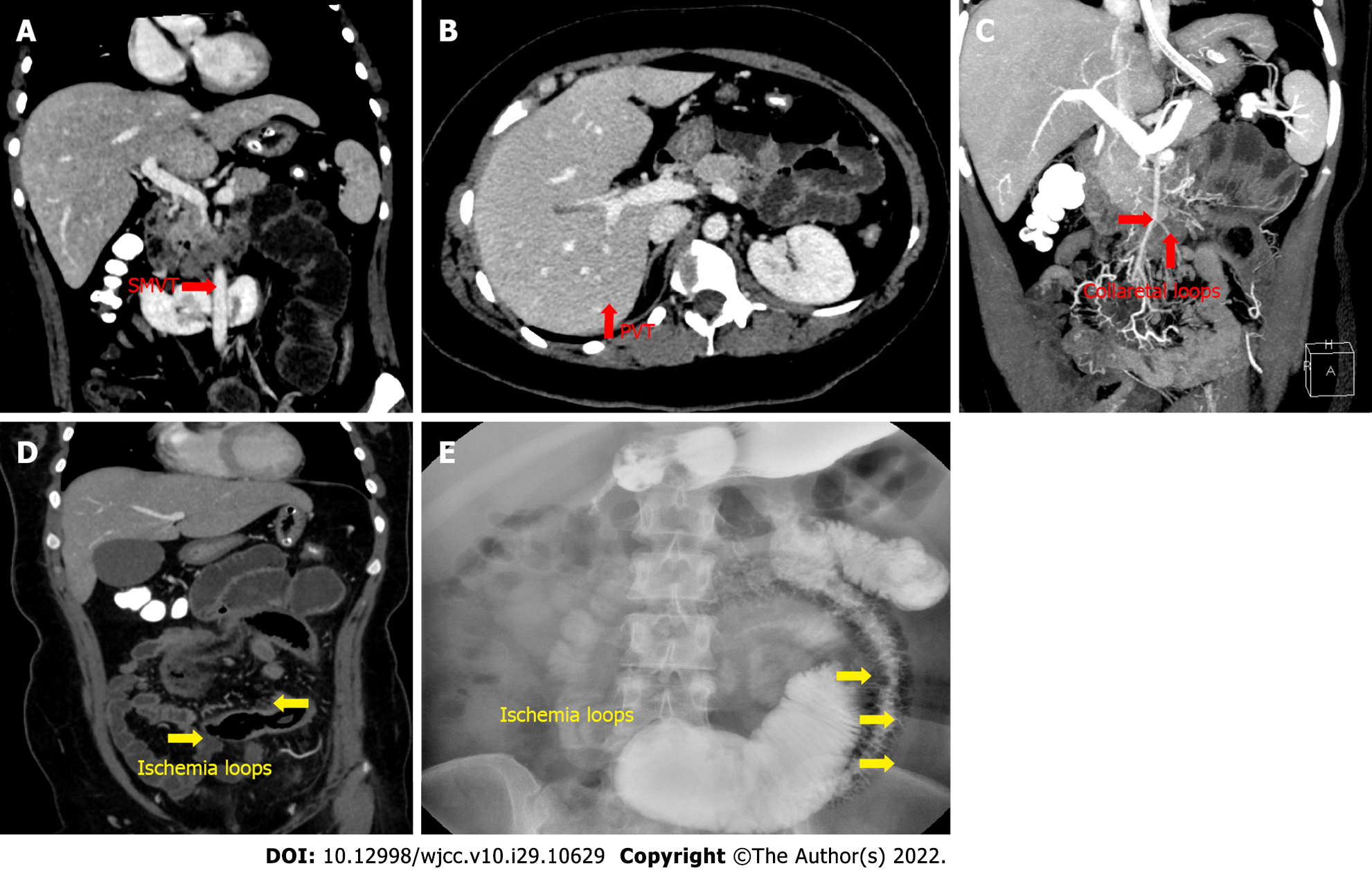
Figure 3 Follow-up contrast-enhanced computed tomography of the abdomen and total gastroenterography on day 15 after admission.
A and B: Residual thrombotic material visible in the superior mesenteric vein (A) and portal vein thrombosis (B) on contrast-enhanced computed tomography (CECT); C: Collateral vessels (red arrows) on CECT; D: Ischemic intestinal loops (yellow arrows) on CECT; E: Total gastroenterography revealed ischemic intestinal loops (yellow arrows). PVT: Portal vein thrombosis; SMVT: Superior mesenteric vein thrombosis.
- Citation: Zhao JW, Cui XH, Zhao WY, Wang L, Xing L, Jiang XY, Gong X, Yu L. Acute mesenteric ischemia secondary to oral contraceptive-induced portomesenteric and splenic vein thrombosis: A case report. World J Clin Cases 2022; 10(29): 10629-10637
- URL: https://www.wjgnet.com/2307-8960/full/v10/i29/10629.htm
- DOI: https://dx.doi.org/10.12998/wjcc.v10.i29.10629


