Copyright
©The Author(s) 2022.
World J Clin Cases. Oct 16, 2022; 10(29): 10622-10628
Published online Oct 16, 2022. doi: 10.12998/wjcc.v10.i29.10622
Published online Oct 16, 2022. doi: 10.12998/wjcc.v10.i29.10622
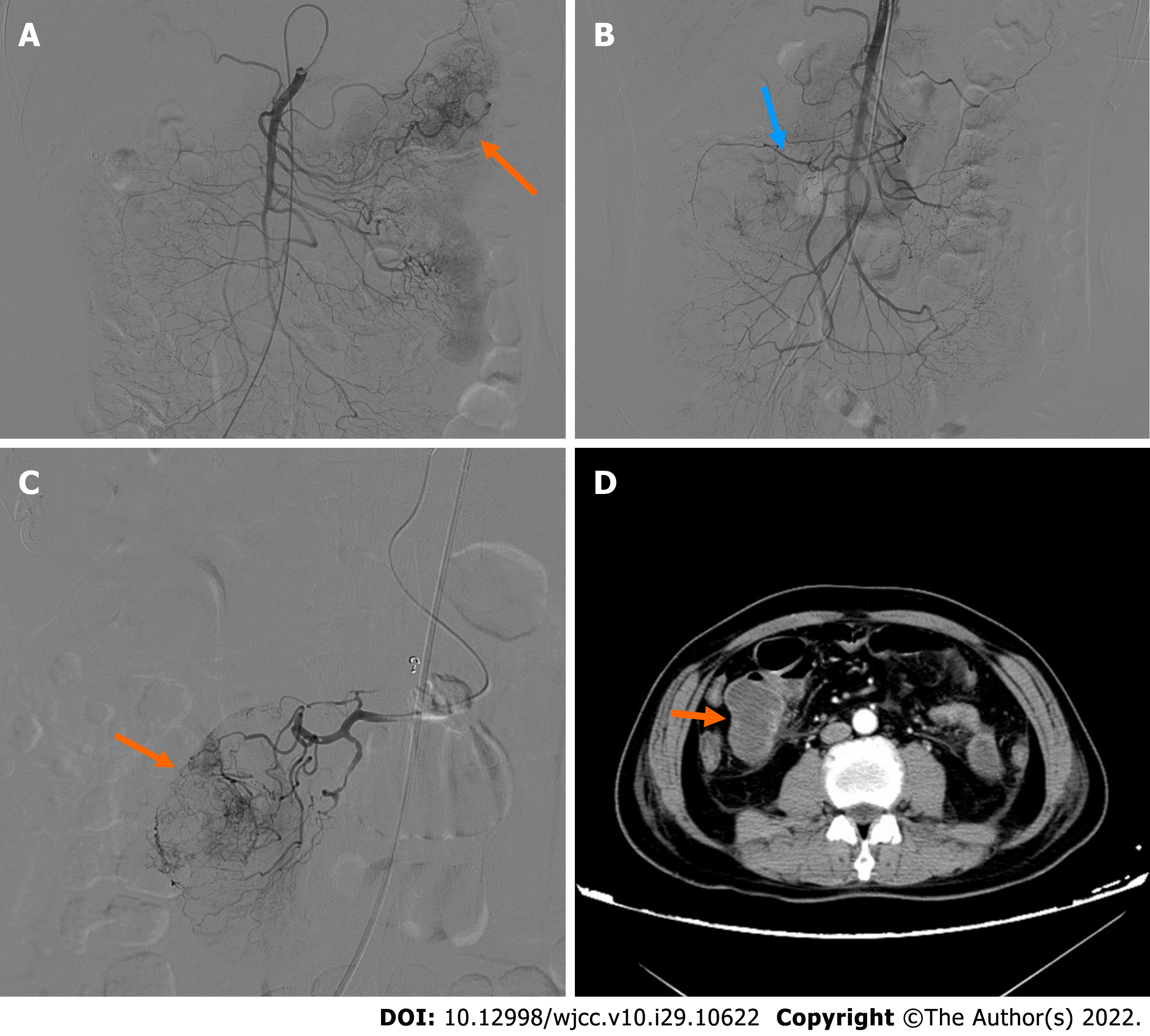
Figure 2 Digital subtraction angiography imaging.
A: First digital subtraction angiography (DSA) showing the mass located in the left abdomen. The distal vascular branches of the first branch of the jejunum appear to be abundant and haphazard with contrast media exudation and retention in the intestinal lumen; B: Second DSA shows the first and second branches of the jejunum in the right abdomen; C: Superselective arteriography showing tumor staining; D: Contrast-enhanced computed tomography after the second embolization showing the mass in the right mid-abdomen.
- Citation: Su JZ, Fan SF, Song X, Cao LJ, Su DY. Wandering small intestinal stromal tumor: A case report. World J Clin Cases 2022; 10(29): 10622-10628
- URL: https://www.wjgnet.com/2307-8960/full/v10/i29/10622.htm
- DOI: https://dx.doi.org/10.12998/wjcc.v10.i29.10622


