Copyright
©The Author(s) 2022.
World J Clin Cases. Oct 16, 2022; 10(29): 10622-10628
Published online Oct 16, 2022. doi: 10.12998/wjcc.v10.i29.10622
Published online Oct 16, 2022. doi: 10.12998/wjcc.v10.i29.10622
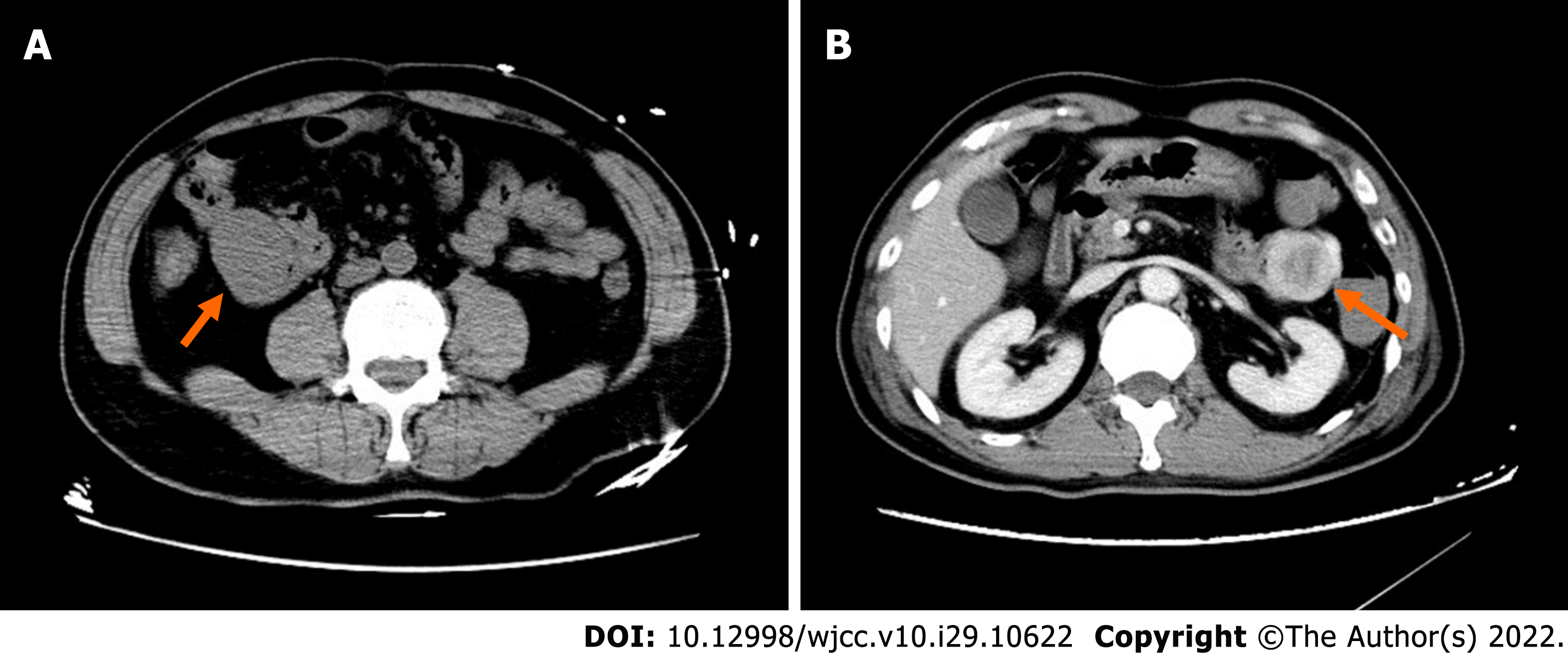
Figure 1 Computed tomography imaging.
A: A 68-year-old man with a wandering small intestinal stromal tumor. At the initial visit, non-contrast computed tomography (CT) scan shows a 4.1 cm × 3.3 cm × 5.0 cm mass located below the right kidney in the right upper abdomen and anterior border of the major lumbar muscle, and the lesion is not clearly demarcated from the small intestine; B: Two days later, contrast-enhanced CT shows the mass (4.6 cm × 3.4 cm × 5.0 cm) in the upper left abdomen in front of the left kidney with non-homogenous enhancement and a thickened artery visible at the edge of the mass. The mass on right upper abdomen cannot be visualized.
- Citation: Su JZ, Fan SF, Song X, Cao LJ, Su DY. Wandering small intestinal stromal tumor: A case report. World J Clin Cases 2022; 10(29): 10622-10628
- URL: https://www.wjgnet.com/2307-8960/full/v10/i29/10622.htm
- DOI: https://dx.doi.org/10.12998/wjcc.v10.i29.10622


