Copyright
©The Author(s) 2022.
World J Clin Cases. Oct 16, 2022; 10(29): 10606-10613
Published online Oct 16, 2022. doi: 10.12998/wjcc.v10.i29.10606
Published online Oct 16, 2022. doi: 10.12998/wjcc.v10.i29.10606
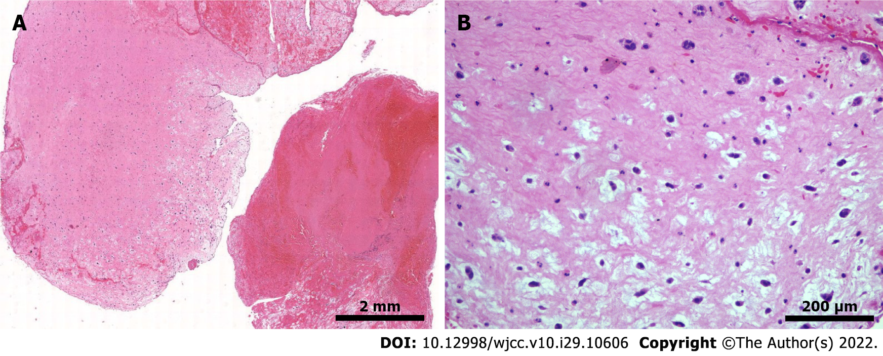
Figure 3 Hematological staining.
A: Histologist section shows embolism with thrombus components and mucous background, with scattered sparse cell clusters and multiple villi-like protrusions, which are consistent with the characteristics of soft and brittle taxonomy (1 ×); B: High magnification microscope shows scattered free clusters on a mucus background with nested round, polygonal cells (squamous cells) arranged in short cords, with round nuclei and fine chromatin, and no atypical (10 ×).
- Citation: Meng XH, Xie LS, Xie XP, Liu YC, Huang CP, Wang LJ, Zhang GH, Xu D, Cai XC, Fang X. Cardiac myxoma shedding leads to lower extremity arterial embolism: A case report. World J Clin Cases 2022; 10(29): 10606-10613
- URL: https://www.wjgnet.com/2307-8960/full/v10/i29/10606.htm
- DOI: https://dx.doi.org/10.12998/wjcc.v10.i29.10606


