Copyright
©The Author(s) 2022.
World J Clin Cases. Oct 6, 2022; 10(28): 10387-10390
Published online Oct 6, 2022. doi: 10.12998/wjcc.v10.i28.10387
Published online Oct 6, 2022. doi: 10.12998/wjcc.v10.i28.10387
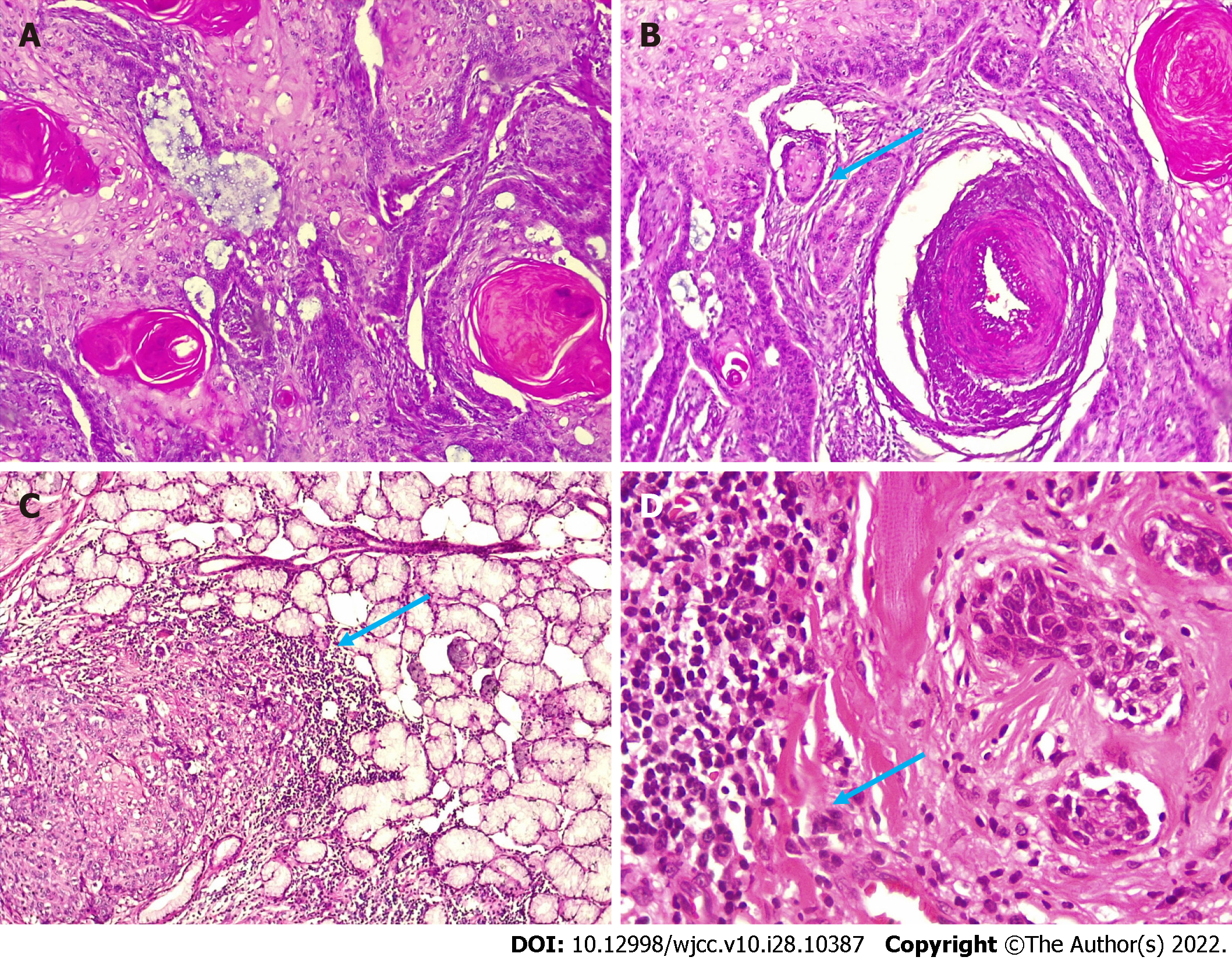
Figure 2 Hematoxylin and eosin staining.
A: Well-differentiated squamous cell carcinoma (hematoxylin and eosin staining, magnification 100 ×); B: Epithelial islands formed by tumor cells (hematoxylin and eosin staining, magnification 100 ×); C: Neoplastic cells infiltrating glandular tissue (hematoxylin and eosin staining, magnification 100 ×); D: Neoplastic cells with an infiltrating pattern in blood vessels (hematoxylin and eosin staining, magnification 400 ×).
- Citation: Cuevas-González JC, Cuevas-González MV, Espinosa-Cristobal LF, Donohue Cornejo A. Tumor invasion front in oral squamous cell carcinoma. World J Clin Cases 2022; 10(28): 10387-10390
- URL: https://www.wjgnet.com/2307-8960/full/v10/i28/10387.htm
- DOI: https://dx.doi.org/10.12998/wjcc.v10.i28.10387


