Copyright
©The Author(s) 2022.
World J Clin Cases. Oct 6, 2022; 10(28): 10358-10365
Published online Oct 6, 2022. doi: 10.12998/wjcc.v10.i28.10358
Published online Oct 6, 2022. doi: 10.12998/wjcc.v10.i28.10358
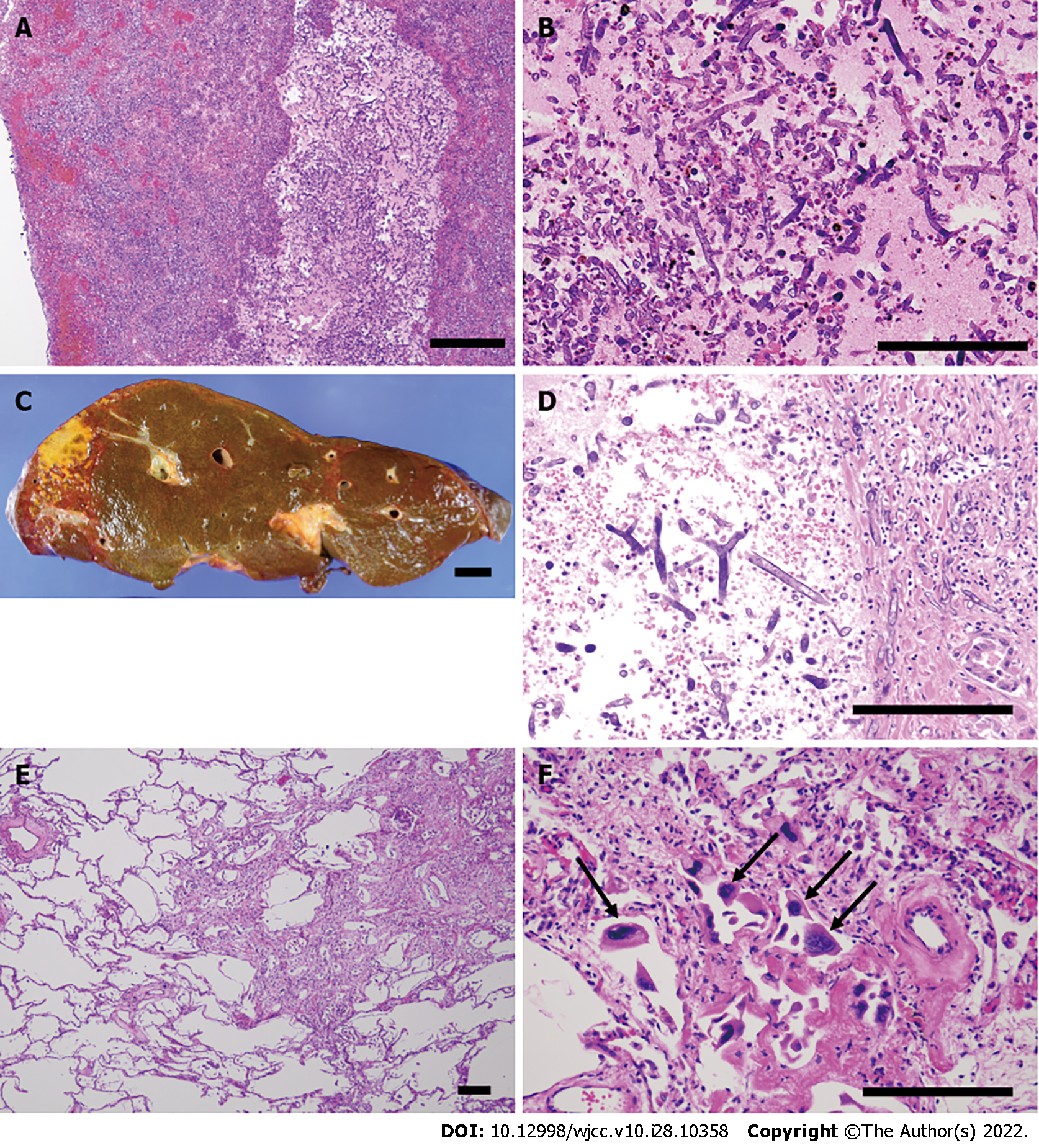
Figure 3 Histopathological findings of the Mucorales infection in the organs and thrombus.
A and B: Presence of Mucorales in the thrombus; H&E staining; magnification × 100 (A), × 200 (B); C: Macroscopic image showing partial necrosis of the liver; D: Hepatic infarction caused by the thrombus including Mucorales; H&E staining; magnification × 200; E: The proliferative/organizing phase of diffuse alveolar damage of the left lung. Restoration of type II pneumocytes and proliferation of myofibroblasts are partially shown. H&E staining; magnification × 40; F: Cytomegalovirus infection. Cytomegalovirus-infected cells are indicated by black arrows. H&E staining; magnification × 200. Bar in the autopsy images: 2 cm; Bar in the microscopic images: 200 μm.
- Citation: Kyuno D, Kubo T, Tsujiwaki M, Sugita S, Hosaka M, Ito H, Harada K, Takasawa A, Kubota Y, Takasawa K, Ono Y, Magara K, Narimatsu E, Hasegawa T, Osanai M. COVID-19-associated disseminated mucormycosis: An autopsy case report. World J Clin Cases 2022; 10(28): 10358-10365
- URL: https://www.wjgnet.com/2307-8960/full/v10/i28/10358.htm
- DOI: https://dx.doi.org/10.12998/wjcc.v10.i28.10358


