Copyright
©The Author(s) 2022.
World J Clin Cases. Oct 6, 2022; 10(28): 10358-10365
Published online Oct 6, 2022. doi: 10.12998/wjcc.v10.i28.10358
Published online Oct 6, 2022. doi: 10.12998/wjcc.v10.i28.10358
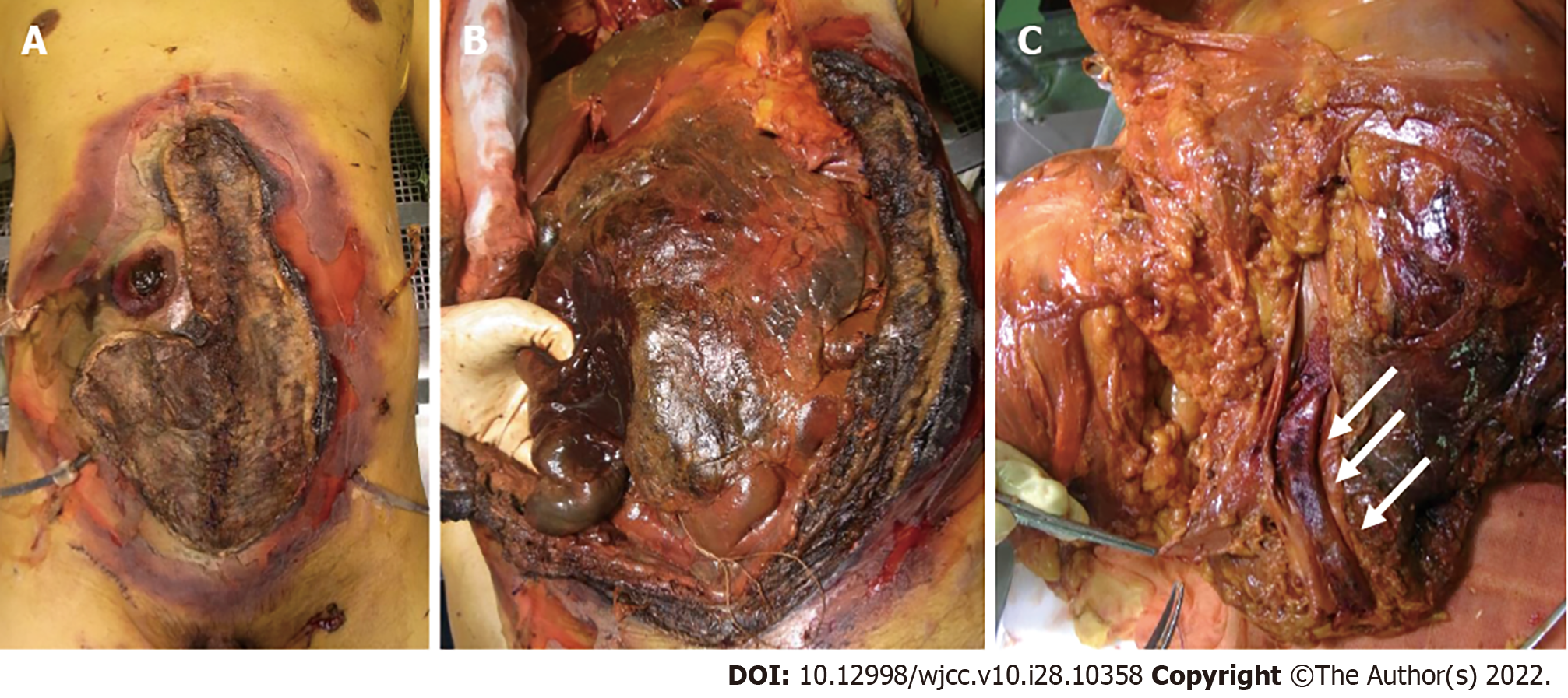
Figure 2 Autopsy findings of the Mucorales infection.
A: The image shows the necrotic stoma, skin, and abdominal wall; B: The necrotic abdominal organs are seen in this image. A pathologist held the intestine on the oral side of the stoma; C: Dorsal view of the incised common iliac vein. The thrombus can be seen in the common iliac vein (indicated by arrows).
- Citation: Kyuno D, Kubo T, Tsujiwaki M, Sugita S, Hosaka M, Ito H, Harada K, Takasawa A, Kubota Y, Takasawa K, Ono Y, Magara K, Narimatsu E, Hasegawa T, Osanai M. COVID-19-associated disseminated mucormycosis: An autopsy case report. World J Clin Cases 2022; 10(28): 10358-10365
- URL: https://www.wjgnet.com/2307-8960/full/v10/i28/10358.htm
- DOI: https://dx.doi.org/10.12998/wjcc.v10.i28.10358


