Copyright
©The Author(s) 2022.
World J Clin Cases. Oct 6, 2022; 10(28): 10358-10365
Published online Oct 6, 2022. doi: 10.12998/wjcc.v10.i28.10358
Published online Oct 6, 2022. doi: 10.12998/wjcc.v10.i28.10358
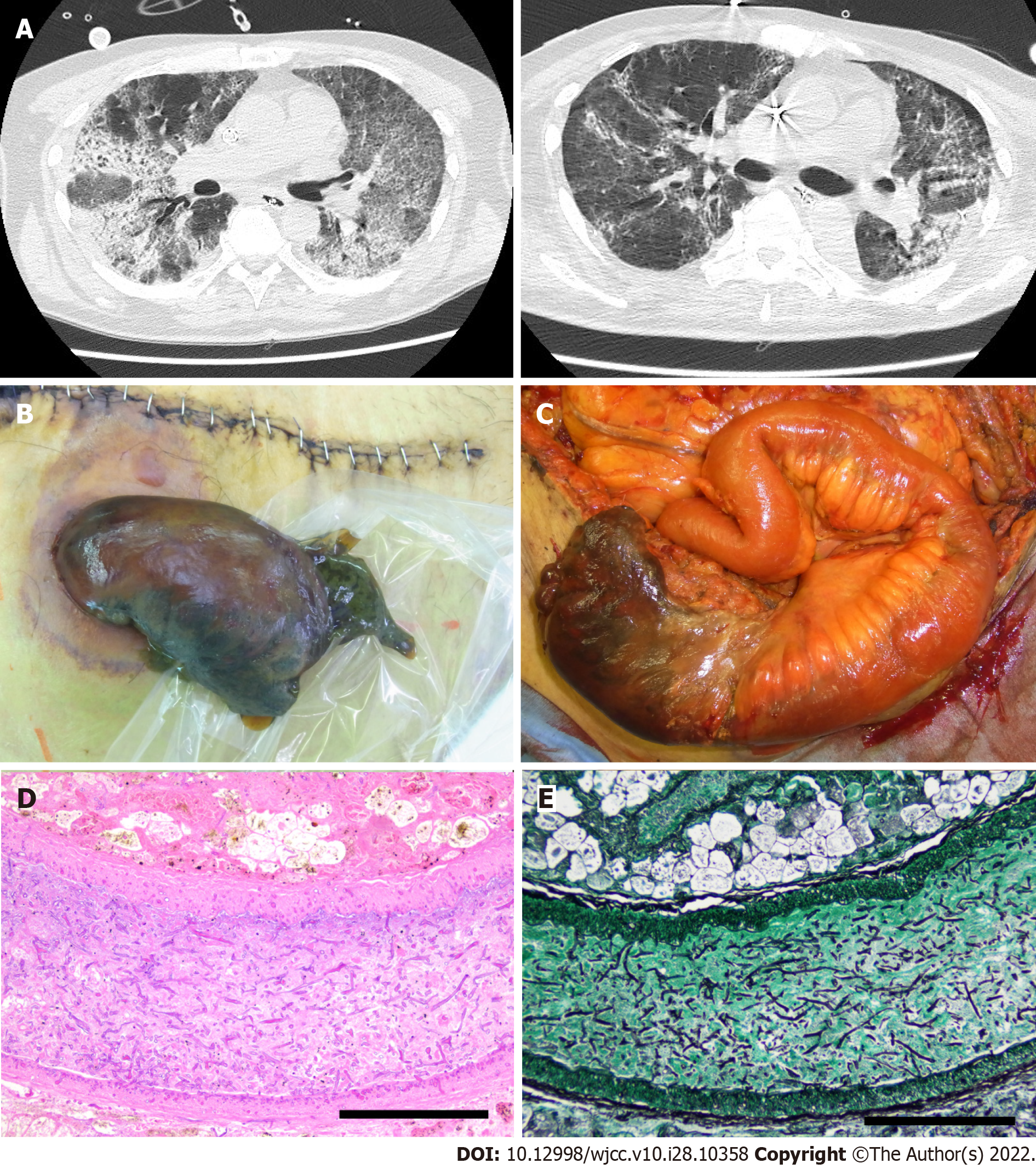
Figure 1 Imaging and histological findings during treatment.
A: Computed tomography (CT) images of acute respiratory distress syndrome (ARDS). Left image: CT image at the time of ARDS diagnosis. Right image: CT image after 38 d; B: The necrotic ileostoma caused by the mucormycosis can be seen in this image; C: Surgical findings at the stomal reconstruction; D and E; Presence of thrombus with Mucorales in the mesenteric vessels of the necrotic ileostoma. H&E staining; magnification × 200 (D), Grocott staining; magnification × 200 (E). Bar: 200 μm.
- Citation: Kyuno D, Kubo T, Tsujiwaki M, Sugita S, Hosaka M, Ito H, Harada K, Takasawa A, Kubota Y, Takasawa K, Ono Y, Magara K, Narimatsu E, Hasegawa T, Osanai M. COVID-19-associated disseminated mucormycosis: An autopsy case report. World J Clin Cases 2022; 10(28): 10358-10365
- URL: https://www.wjgnet.com/2307-8960/full/v10/i28/10358.htm
- DOI: https://dx.doi.org/10.12998/wjcc.v10.i28.10358


