Copyright
©The Author(s) 2022.
World J Clin Cases. Oct 6, 2022; 10(28): 10346-10357
Published online Oct 6, 2022. doi: 10.12998/wjcc.v10.i28.10346
Published online Oct 6, 2022. doi: 10.12998/wjcc.v10.i28.10346
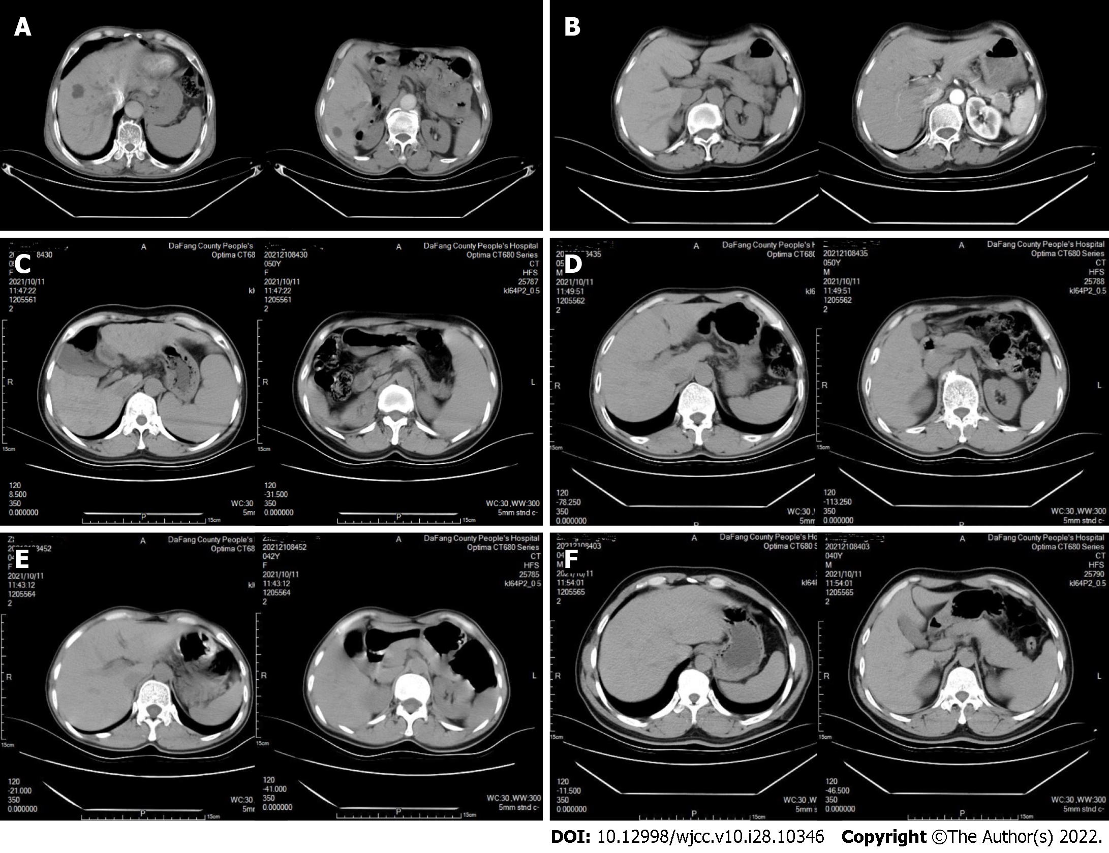
Figure 3 Computed tomography images of the upper abdomens of six family members.
A and B: I-1, I-2 (2022 Jan 20): The size and shape of liver and spleen were normal, and there was no obvious enhancement on enhanced scan; C: II-2 (2021 Oct 11): The proportions of the liver lobes were disordered and the contour was not regular; multiple low-density shadows were on the boundary, and elliptic shapes with different sizes were observed in liver, with a computed tomography (CT) value of about 13 HU; the spleen volume was significantly increased and the parenchyma density was uniform; there was no fluid accumulation in the abdominal cavity; D-F: II-3, II-4, and II-5 (2021 Oct 11): The liver was normal in size and shape had a smooth surface and a uniform density of parenchyma; the spleen was normal in size and density.
- Citation: Jiang JL, Qian JF, Xiao DH, Liu X, Zhu F, Wang J, Xing ZX, Xu DL, Xue Y, He YH. Relationship of familial cytochrome P450 4V2 gene mutation with liver cirrhosis: A case report and review of the literature. World J Clin Cases 2022; 10(28): 10346-10357
- URL: https://www.wjgnet.com/2307-8960/full/v10/i28/10346.htm
- DOI: https://dx.doi.org/10.12998/wjcc.v10.i28.10346


