Copyright
©The Author(s) 2022.
World J Clin Cases. Oct 6, 2022; 10(28): 10346-10357
Published online Oct 6, 2022. doi: 10.12998/wjcc.v10.i28.10346
Published online Oct 6, 2022. doi: 10.12998/wjcc.v10.i28.10346
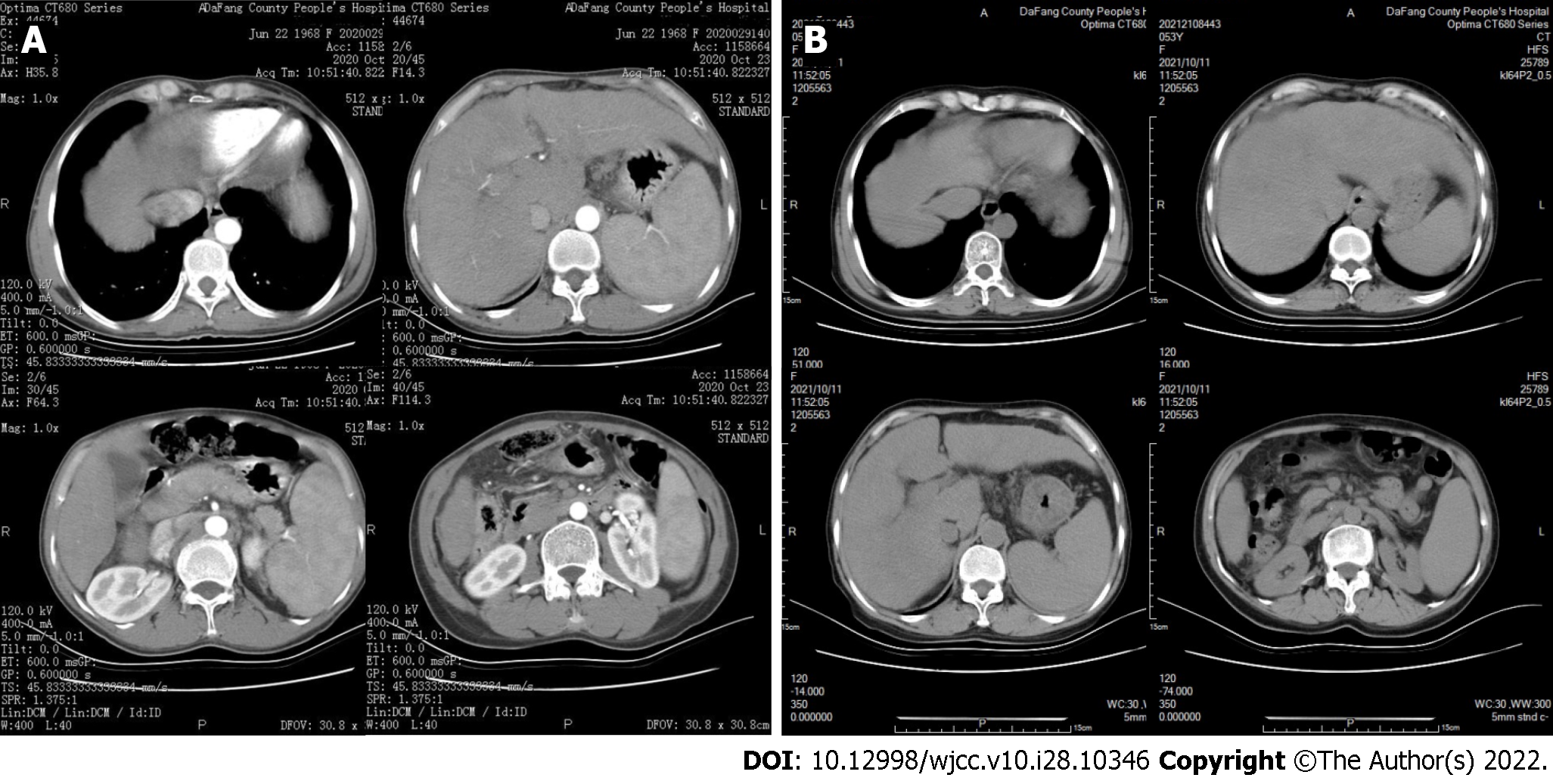
Figure 1 Computed tomography images of the upper abdomen of the index patient (II-1).
A: Enhanced computed tomography (CT) (2020 Oct 23): The proportions of liver were disordered, the contour was not regular, and there was no obvious enhancement of liver parenchyma; the spleen was enlarged; a fluid density shadow was evident in the abdominal cavity; B: Scanning CT (2021 Oct 11): A fluid density shadow was present at the edge of liver; liver volume was increased; a liver fissure was widened, with uneven and blunt edges and no abnormal density foci; the spleen was enlarged and parenchyma density was uniform.
- Citation: Jiang JL, Qian JF, Xiao DH, Liu X, Zhu F, Wang J, Xing ZX, Xu DL, Xue Y, He YH. Relationship of familial cytochrome P450 4V2 gene mutation with liver cirrhosis: A case report and review of the literature. World J Clin Cases 2022; 10(28): 10346-10357
- URL: https://www.wjgnet.com/2307-8960/full/v10/i28/10346.htm
- DOI: https://dx.doi.org/10.12998/wjcc.v10.i28.10346


