Copyright
©The Author(s) 2022.
World J Clin Cases. Oct 6, 2022; 10(28): 10317-10325
Published online Oct 6, 2022. doi: 10.12998/wjcc.v10.i28.10317
Published online Oct 6, 2022. doi: 10.12998/wjcc.v10.i28.10317
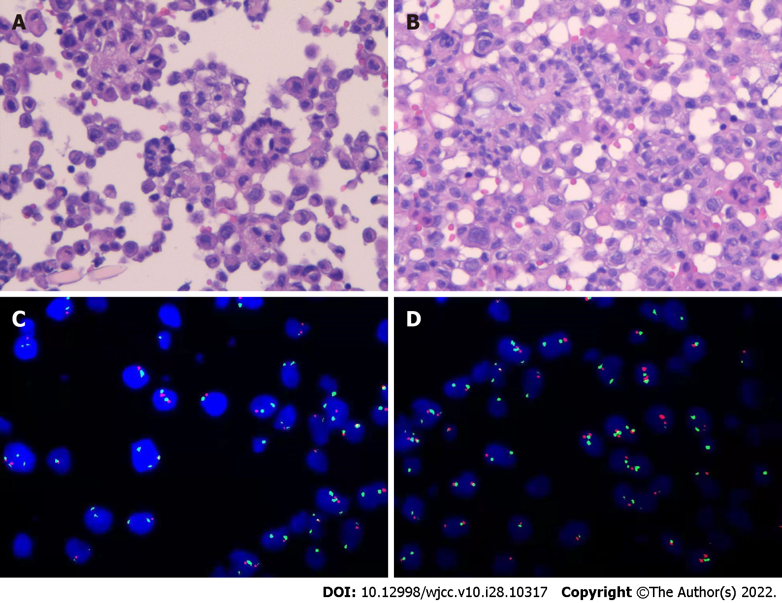
Figure 1 Histopathological and fluorescence in situ hybridization features.
A and B: A large number of heterozygous cells, some of which are papillary structures, increased nucleocytoplasmic ratio, hyperchromatic nuclei, and obvious atypia (hematoxylin and eosin stain; scale bar: 100 μm); C and D: Fluorescence in situ hybridization detection results: CDKN2A (P16) probe (+), number of missing cells/total counted cells = 41%.
- Citation: Huang X, Hong Y, Xie SY, Liao HL, Huang HM, Liu JH, Long WJ. Malignant peritoneal mesothelioma with massive ascites as the first symptom: A case report. World J Clin Cases 2022; 10(28): 10317-10325
- URL: https://www.wjgnet.com/2307-8960/full/v10/i28/10317.htm
- DOI: https://dx.doi.org/10.12998/wjcc.v10.i28.10317


