Copyright
©The Author(s) 2022.
World J Clin Cases. Oct 6, 2022; 10(28): 10301-10309
Published online Oct 6, 2022. doi: 10.12998/wjcc.v10.i28.10301
Published online Oct 6, 2022. doi: 10.12998/wjcc.v10.i28.10301
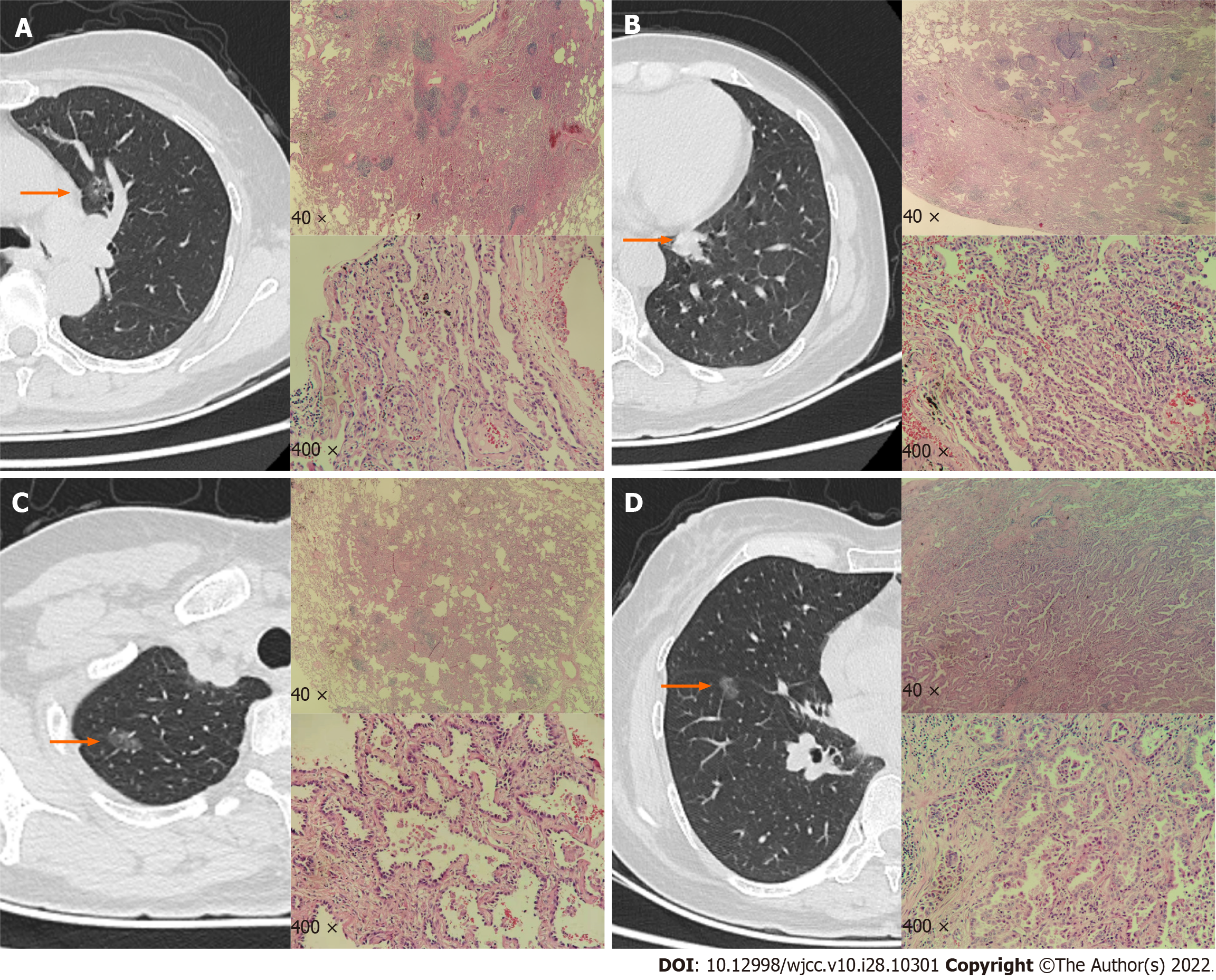
Figure 3 The left side of the image is a computed tomography image.
The red arrow indicates the location of the nodule. The upper right side indicates a low-power microscopic pathological image, and the lower right side indicates a high-power microscopic pathological image. A: The left upper pulmonary nodule is an adenocarcinoma in situ, with adherent growth and obvious cell atypia; B: The left lower lung nodule is an acinar adenocarcinoma with glandular arrangement, invasive growth, significant atypia of cancer cells, and proliferation of interstitial fibrous tissue; C and D: The right upper and right lower nodules are microinvasive adenocarcinoma. The cancer cells adhere to the wall; the cell atypia is significant; and the focus shows invasive growth.
- Citation: Zhang DY, Liu J, Zhang Y, Ye JY, Hu S, Zhang WX, Yu DL, Wei YP. One-stage resection of four genotypes of bilateral multiple primary lung adenocarcinoma: A case report. World J Clin Cases 2022; 10(28): 10301-10309
- URL: https://www.wjgnet.com/2307-8960/full/v10/i28/10301.htm
- DOI: https://dx.doi.org/10.12998/wjcc.v10.i28.10301


