Copyright
©The Author(s) 2022.
World J Clin Cases. Oct 6, 2022; 10(28): 10252-10259
Published online Oct 6, 2022. doi: 10.12998/wjcc.v10.i28.10252
Published online Oct 6, 2022. doi: 10.12998/wjcc.v10.i28.10252
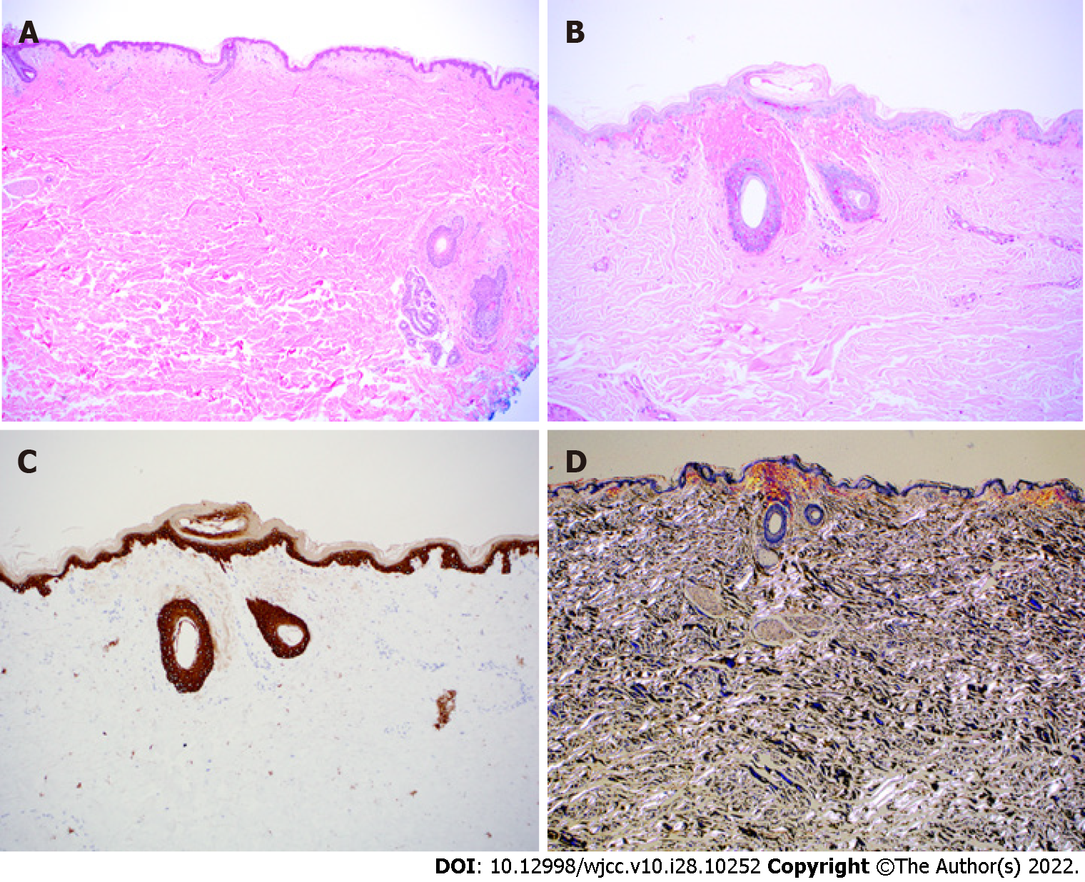
Figure 3 Histopathology of abdominal fat pad biopsy.
A: H&E stain at 4 × of skin biopsy with eosinophilic deposits in the upper papillary dermis and periadnexal dermis; B: Periodic Acid-Schiff stain at 10 × shows the deposition to be negative for lipoid proteinosis; C: Cytokeratin 5/6 immunostain at 10 × is blush positive but interpreted as negative, an important stain considering differential includes primary cutaneous amyloidosis of macular or lichen subtype; D: Congo red stain at 4 × under polarization showing apple green birefringence, rarely seen in cutaneous amyloidosis but always present in amyloid light amyloidosis.
- Citation: Bilton SE, Shah N, Dougherty D, Simpson S, Holliday A, Sahebjam F, Grider DJ. Persistent diarrhea with petechial rash - unusual pattern of light chain amyloidosis deposition on skin and gastrointestinal biopsies: A case report. World J Clin Cases 2022; 10(28): 10252-10259
- URL: https://www.wjgnet.com/2307-8960/full/v10/i28/10252.htm
- DOI: https://dx.doi.org/10.12998/wjcc.v10.i28.10252


