Copyright
©The Author(s) 2022.
World J Clin Cases. Oct 6, 2022; 10(28): 10244-10251
Published online Oct 6, 2022. doi: 10.12998/wjcc.v10.i28.10244
Published online Oct 6, 2022. doi: 10.12998/wjcc.v10.i28.10244
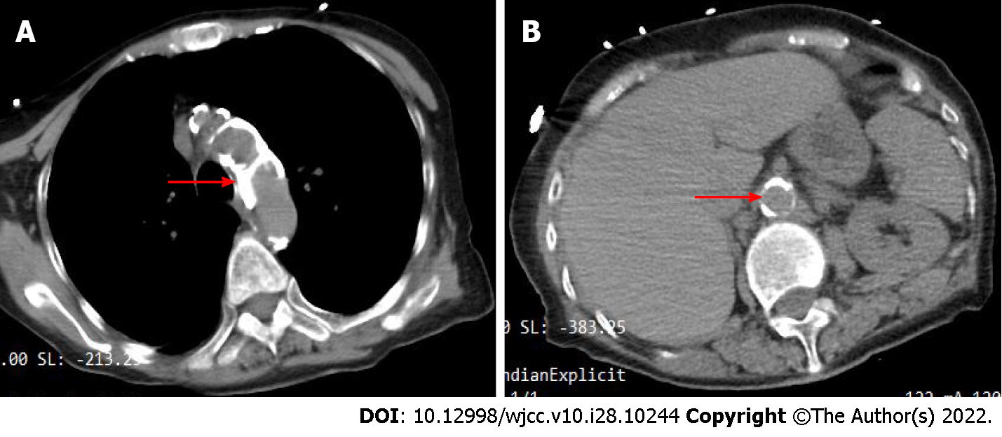
Figure 3 Abdominal computed tomography findings.
Chest and abdominal computed tomography images of the patient at admission when the patient was admitted to the hospital (November 16, 2021). A: The patient's aortic arch has markedly extensive calcification (red arrow); B: The patient's abdominal aorta has obvious calcification (red arrow indicates the opening of the superior mesenteric artery).
- Citation: Ding P, Zhou Y, Long KL, Zhang S, Gao PY. Acute mesenteric ischemia due to percutaneous coronary intervention: A case report. World J Clin Cases 2022; 10(28): 10244-10251
- URL: https://www.wjgnet.com/2307-8960/full/v10/i28/10244.htm
- DOI: https://dx.doi.org/10.12998/wjcc.v10.i28.10244


