Copyright
©The Author(s) 2022.
World J Clin Cases. Oct 6, 2022; 10(28): 10244-10251
Published online Oct 6, 2022. doi: 10.12998/wjcc.v10.i28.10244
Published online Oct 6, 2022. doi: 10.12998/wjcc.v10.i28.10244
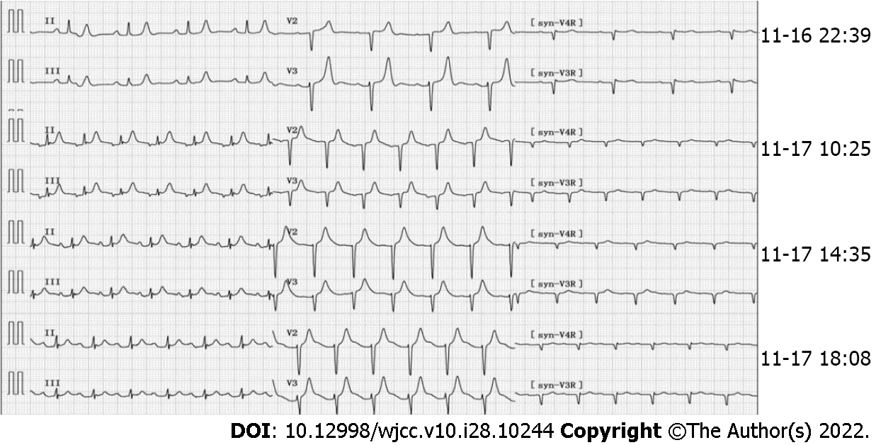
Figure 2 Electrocardiogram findings at different time points.
The initial electrocardiogram (ECG) (November 16, 2021, 22:39): Sinus rhythm, high and sharp T wave; R wave: V1-V4 progression was poor, V1 was Qr type. ECG (November 17, 2021, 10:25): Sinus tachycardia, paroxysmal atrial tachycardia, atrial fusion waves, and ST segment elevation in leads II, III, and avF was 0.10mV. ECGs (November 17, 2021, 14:35 and 18:08) reported ST-segment elevation of II, III, and avF exceeding 0.05 mV.
- Citation: Ding P, Zhou Y, Long KL, Zhang S, Gao PY. Acute mesenteric ischemia due to percutaneous coronary intervention: A case report. World J Clin Cases 2022; 10(28): 10244-10251
- URL: https://www.wjgnet.com/2307-8960/full/v10/i28/10244.htm
- DOI: https://dx.doi.org/10.12998/wjcc.v10.i28.10244


