Copyright
©The Author(s) 2022.
World J Clin Cases. Oct 6, 2022; 10(28): 10201-10207
Published online Oct 6, 2022. doi: 10.12998/wjcc.v10.i28.10201
Published online Oct 6, 2022. doi: 10.12998/wjcc.v10.i28.10201
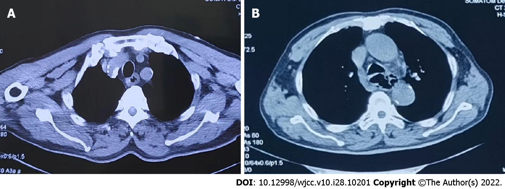
Figure 2 Computed tomography images of the lesion.
A: Upon admission showing artery calcification, thickened oesophageal wall, and a dilated lumen; B: On day 7 showing thickened, dilated, and irregularly shaped oesophageal wall.
- Citation: Li YQ, Yu GC, Shi LK, Zhao LW, Wen ZX, Kan BT, Jian XD. Clinical analysis of pipeline dredging agent poisoning: A case report. World J Clin Cases 2022; 10(28): 10201-10207
- URL: https://www.wjgnet.com/2307-8960/full/v10/i28/10201.htm
- DOI: https://dx.doi.org/10.12998/wjcc.v10.i28.10201


