Copyright
©The Author(s) 2022.
World J Clin Cases. Oct 6, 2022; 10(28): 10130-10135
Published online Oct 6, 2022. doi: 10.12998/wjcc.v10.i28.10130
Published online Oct 6, 2022. doi: 10.12998/wjcc.v10.i28.10130
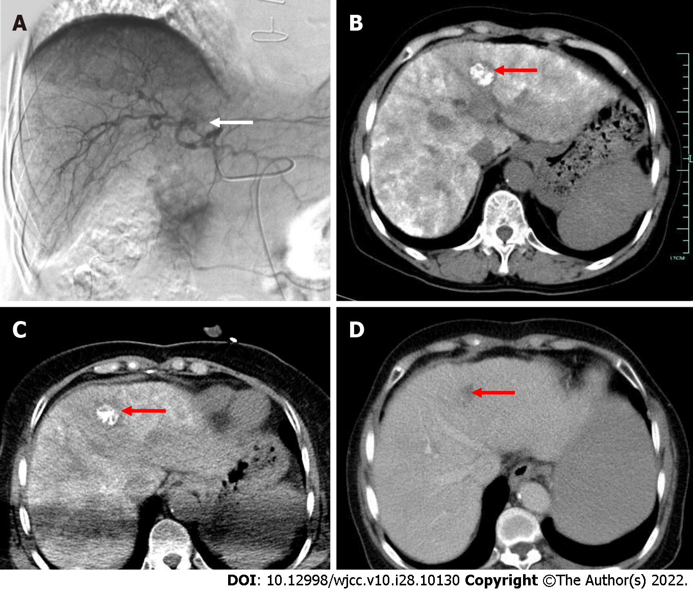
Figure 2 Patient’s digital subtraction angiography and computed tomography images.
A: Hepatic arteriography, transcatheter arterial chemoembolization (TACE), and hepatic artery digital subtraction angiography imaging showed abnormal staining (white arrow) on the left hepatic artery on January 29, 2015; B: Dense lipiodol deposition in the left lobe lesions was observed two days after TACE by computed tomography (CT) scanning (red arrow); C: CT-guided percutaneous radiofrequency ablation was performed on the hepatic lesion (red arrow); D: One month after the procedure, the tumour had regressed completely (red arrow).
- Citation: Wu FZ, Chen XX, Chen WY, Wu QH, Mao JT, Zhao ZW. Multiple primary malignancies – hepatocellular carcinoma combined with splenic lymphoma: A case report. World J Clin Cases 2022; 10(28): 10130-10135
- URL: https://www.wjgnet.com/2307-8960/full/v10/i28/10130.htm
- DOI: https://dx.doi.org/10.12998/wjcc.v10.i28.10130


