Copyright
©The Author(s) 2022.
World J Clin Cases. Oct 6, 2022; 10(28): 10130-10135
Published online Oct 6, 2022. doi: 10.12998/wjcc.v10.i28.10130
Published online Oct 6, 2022. doi: 10.12998/wjcc.v10.i28.10130
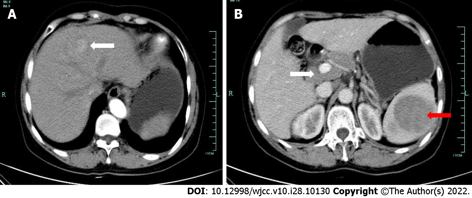
Figure 1 Patient’s enhanced computed tomography images.
A: Enhanced computed tomography examination showed a mass with a diameter of approximately 2.6 cm on the left liver lobe on January 13, 2015; B: The splenic portal phase showed multiple circular low-density shadow areas with a maximum diameter of approximately 7.6 cm (red arrow); multiple enlarged lymph nodes were detected around the liver helium and retroperitoneum with partial fusion conglobation (white arrow).
- Citation: Wu FZ, Chen XX, Chen WY, Wu QH, Mao JT, Zhao ZW. Multiple primary malignancies – hepatocellular carcinoma combined with splenic lymphoma: A case report. World J Clin Cases 2022; 10(28): 10130-10135
- URL: https://www.wjgnet.com/2307-8960/full/v10/i28/10130.htm
- DOI: https://dx.doi.org/10.12998/wjcc.v10.i28.10130


