Copyright
©The Author(s) 2022.
World J Clin Cases. Oct 6, 2022; 10(28): 10120-10129
Published online Oct 6, 2022. doi: 10.12998/wjcc.v10.i28.10120
Published online Oct 6, 2022. doi: 10.12998/wjcc.v10.i28.10120
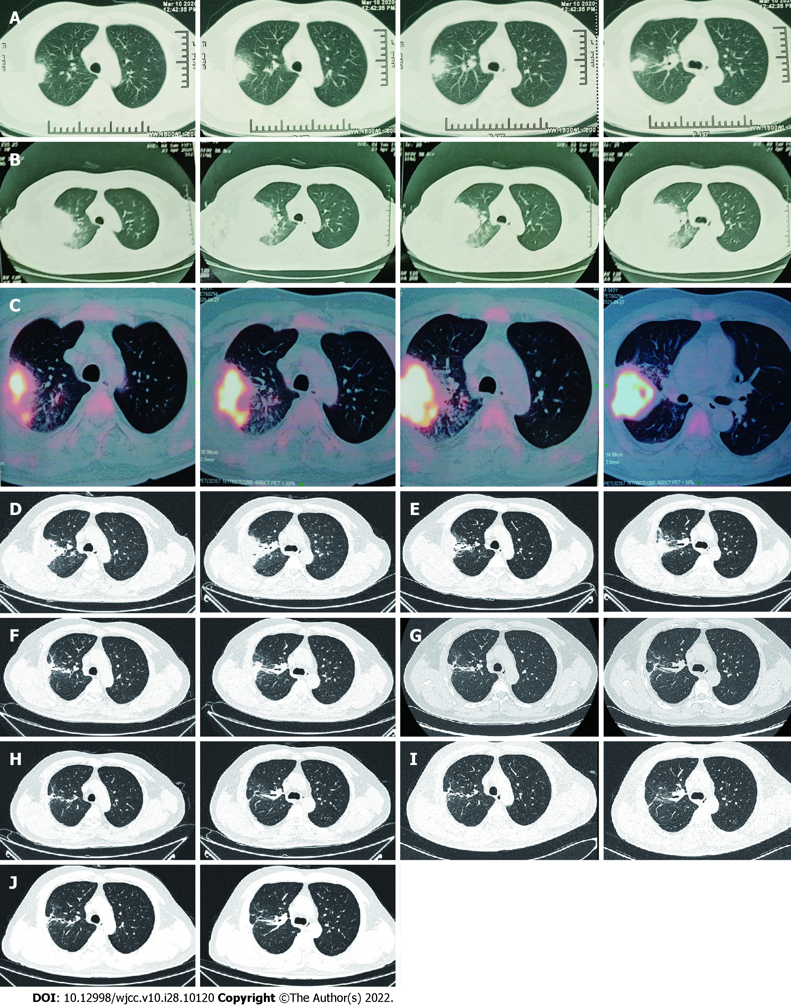
Figure 1 Computed tomography of right lung.
A: The first scan demonstrated a mass and atelectasis in the right upper lobe on March 20, 2020; B: Chest Computed Tomography (CT) showed aggravation of the original lesion on April 23, 2020; C: Positron emission tomography-CT showed that the mass lesions in the posterior segment of the right upper lobe had significantly increased glucose metabolism inhomogeneously on April 27, 2020; D: Primary lesion of the right upper lobe reduced on May 23, 2020; E-J: Repeated pulmonary CT showed primary lesion size of the right upper lobe continued to decrease on different times, involving June 22, 2020 (E), July 29, 2020 (F), September 8, 2020 (G), November 20, 2020 (H), January 7, 2021 (I) and March 25, 2021 (J). CT: Computed tomography.
- Citation: Li T, Chen YX, Lin JJ, Lin WX, Zhang WZ, Dong HM, Cai SX, Meng Y. Successful treatment of disseminated nocardiosis diagnosed by metagenomic next-generation sequencing: A case report and review of literature. World J Clin Cases 2022; 10(28): 10120-10129
- URL: https://www.wjgnet.com/2307-8960/full/v10/i28/10120.htm
- DOI: https://dx.doi.org/10.12998/wjcc.v10.i28.10120


