Copyright
©The Author(s) 2022.
World J Clin Cases. Sep 26, 2022; 10(27): 9954-9960
Published online Sep 26, 2022. doi: 10.12998/wjcc.v10.i27.9954
Published online Sep 26, 2022. doi: 10.12998/wjcc.v10.i27.9954
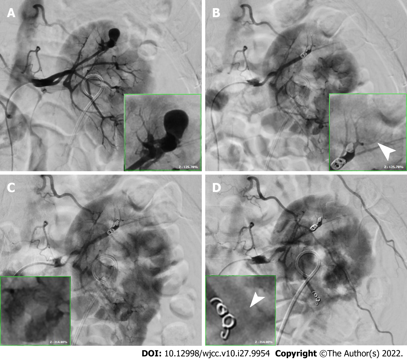
Figure 3 Comparing the image after TAE with residual J-J stent.
A: A 1.5-cm pseudoaneurysm at the upper renal artery branch; B: Embolized using two 0.3 cm × 2 cm and two 0.4 × 2 cm microcoils; C: A 0.4-cm pseudoaneurysm at the lower renal artery terminal branch; D: Embolized using two 0.3 cm × 2 cm microcoils with no contrast extravasation in both lesions (point with white arrowheads).
- Citation: Li YH, Lin YS, Hsu CY, Ou YC, Tung MC. Renal pseudoaneurysm after rigid ureteroscopic lithotripsy: A case report. World J Clin Cases 2022; 10(27): 9954-9960
- URL: https://www.wjgnet.com/2307-8960/full/v10/i27/9954.htm
- DOI: https://dx.doi.org/10.12998/wjcc.v10.i27.9954


