Copyright
©The Author(s) 2022.
World J Clin Cases. Sep 26, 2022; 10(27): 9897-9903
Published online Sep 26, 2022. doi: 10.12998/wjcc.v10.i27.9897
Published online Sep 26, 2022. doi: 10.12998/wjcc.v10.i27.9897
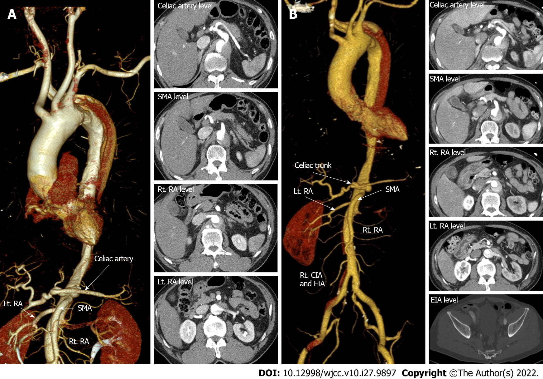
Figure 2 Initial computed tomography post transradial intervention and follow-up computed tomography in 2 days.
A: Emergency computed tomography (CT) aortography immediately after right transradial intervention showing type B aortic dissection (AD) originating from the aberrant right subclavian artery with extension of the intimal flap down the descending to the infrarenal abdominal aorta. The dissection extended into the left renal artery (RA) (red arrowheads in A); B: After two days of intensive care unit stay, follow-up CT showed downstream propagation of the AD into the external iliac artery and left RA with compromised flow (red arrowheads of B). Other arteries including the celiac trunk, superior mesenteric artery and right RA were intact (white arrows) on both CTs. SMA: Superior mesenteric artery; RA: Renal artery; EIA: External iliac artery; CIA: Common iliac artery.
- Citation: Ha K, Jang AY, Shin YH, Lee J, Seo J, Lee SI, Kang WC, Suh SY. Iatrogenic aortic dissection during right transradial intervention in a patient with aberrant right subclavian artery: A case report. World J Clin Cases 2022; 10(27): 9897-9903
- URL: https://www.wjgnet.com/2307-8960/full/v10/i27/9897.htm
- DOI: https://dx.doi.org/10.12998/wjcc.v10.i27.9897


