Copyright
©The Author(s) 2022.
World J Clin Cases. Sep 26, 2022; 10(27): 9873-9878
Published online Sep 26, 2022. doi: 10.12998/wjcc.v10.i27.9873
Published online Sep 26, 2022. doi: 10.12998/wjcc.v10.i27.9873
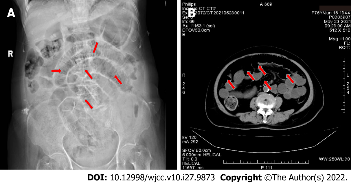
Figure 2 Imaging before treatment.
A: Abdominal X-ray before treatment. Supine position. No definite obstruction point, red arrows indicate dilated small bowel; B: Abdominal computed tomography scan: Small bowel obstruction, no definite obstruction point, red arrows indicate dilated small bowel.
- Citation: Lin YC, Cui XG, Wu LZ, Zhou DQ, Zhou Q. Resolution of herpes zoster-induced small bowel pseudo-obstruction by epidural nerve block: A case report. World J Clin Cases 2022; 10(27): 9873-9878
- URL: https://www.wjgnet.com/2307-8960/full/v10/i27/9873.htm
- DOI: https://dx.doi.org/10.12998/wjcc.v10.i27.9873


