Copyright
©The Author(s) 2022.
World J Clin Cases. Sep 26, 2022; 10(27): 9851-9858
Published online Sep 26, 2022. doi: 10.12998/wjcc.v10.i27.9851
Published online Sep 26, 2022. doi: 10.12998/wjcc.v10.i27.9851
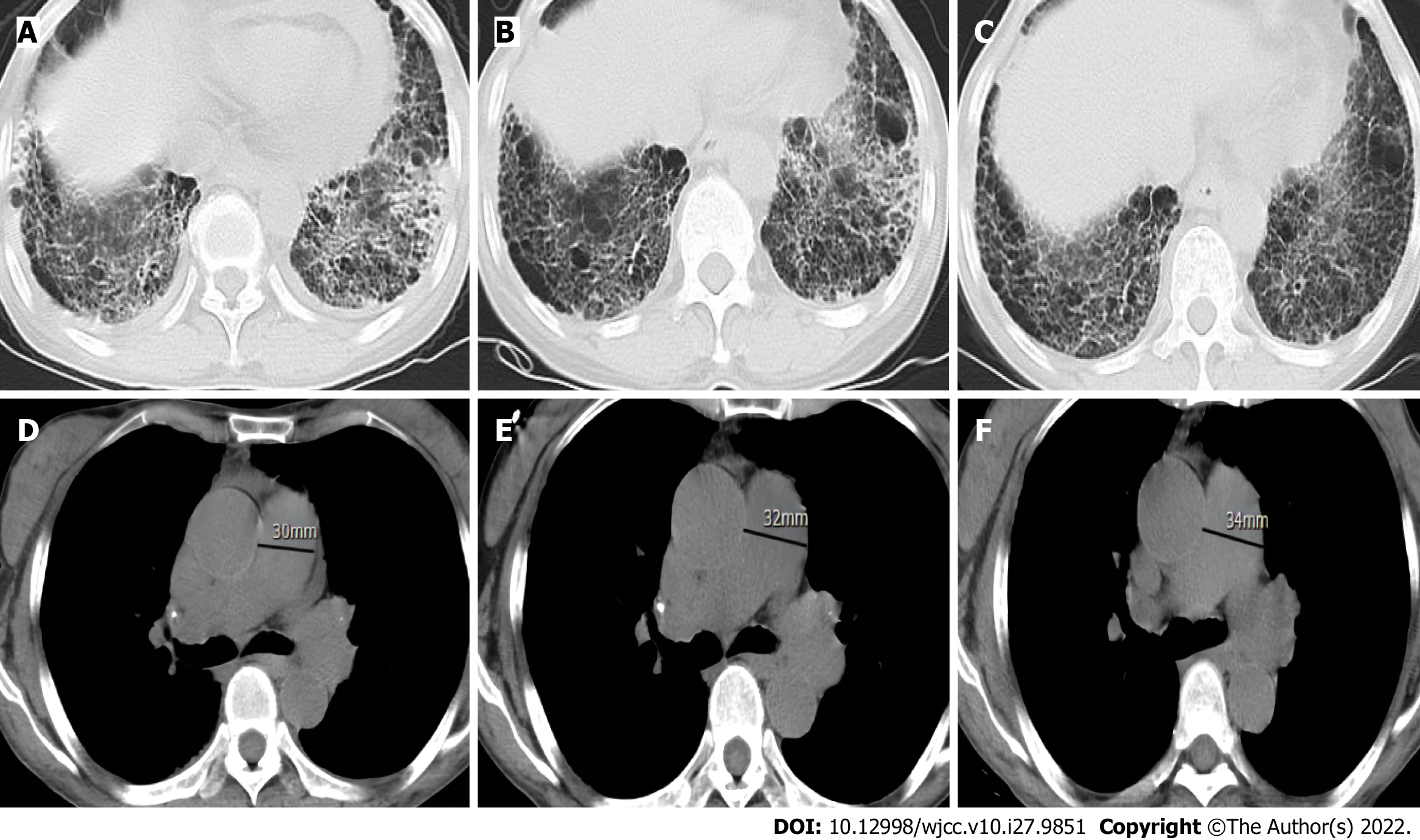
Figure 2 Computed tomography scans over 3 years.
A-C: Chest computed tomography (CT) shows the thickening of both lungs, a subpleural and lower lung mesh texture, and multiple patchy, blurred shadows in the lower lobe of both lungs; the left to right contrast showed no significant progression over 3 years; D-F: Chest CT mediastinal window, with contrast from left to right, showing gradual widening of the pulmonary artery over 3 year.
- Citation: Huang CY, Lu MJ, Tian JH, Liu DS, Wu CY. Pulmonary hypertension secondary to seronegative rheumatoid arthritis overlapping antisynthetase syndrome: A case report. World J Clin Cases 2022; 10(27): 9851-9858
- URL: https://www.wjgnet.com/2307-8960/full/v10/i27/9851.htm
- DOI: https://dx.doi.org/10.12998/wjcc.v10.i27.9851


