Copyright
©The Author(s) 2022.
World J Clin Cases. Sep 26, 2022; 10(27): 9821-9827
Published online Sep 26, 2022. doi: 10.12998/wjcc.v10.i27.9821
Published online Sep 26, 2022. doi: 10.12998/wjcc.v10.i27.9821
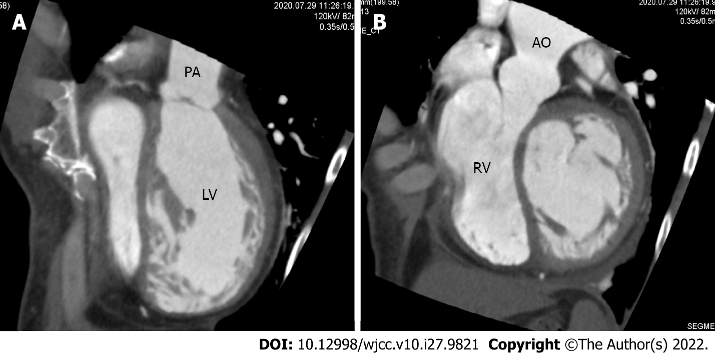
Figure 4 Contrast-enhanced chest computed tomography.
A: The aorta, with the shape of a rough adductor muscle, started from the anatomical right ventricle; B: The pulmonary artery started from the anatomical left ventricle. PA: Pulmonary artery; LV: Left ventricle; RV: Right ventricle; AO: Aorta.
- Citation: Ichii N, Kakinuma T, Fujikawa A, Takeda M, Ohta T, Kagimoto M, Kaneko A, Izumi R, Kakinuma K, Saito K, Maeyama A, Yanagida K, Takeshima N, Ohwada M. Diagnosed corrected transposition of great arteries after cesarean section: A case report. World J Clin Cases 2022; 10(27): 9821-9827
- URL: https://www.wjgnet.com/2307-8960/full/v10/i27/9821.htm
- DOI: https://dx.doi.org/10.12998/wjcc.v10.i27.9821


