Copyright
©The Author(s) 2022.
World J Clin Cases. Sep 26, 2022; 10(27): 9805-9813
Published online Sep 26, 2022. doi: 10.12998/wjcc.v10.i27.9805
Published online Sep 26, 2022. doi: 10.12998/wjcc.v10.i27.9805
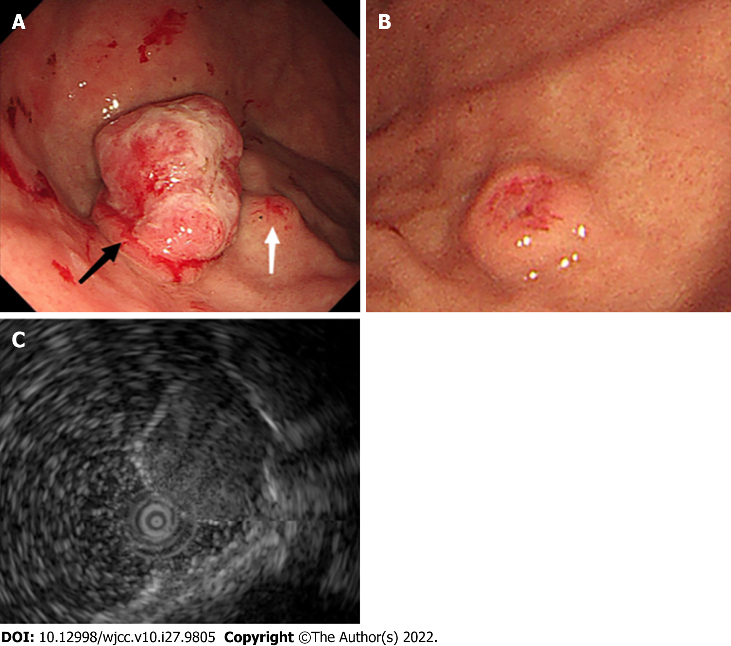
Figure 2 Endoscopic ultrasonography examination in our hospital.
A: A large protruding lesion was found in the fundus, the primary discoid-shape remained in the basal layer of the lesion (black arrow). A small submucosal lesion was detected in the fundus adjacent to the large lesion (white arrow); B: The other similar small submucosal lesion in the middle section of the stomach body; and C: EUS showed a heterogeneous mass that involved the mucosa and submucosal layer, with hypoechoic changes.
- Citation: Chen WG, Shan GD, Zhu HT, Chen LH, Xu GQ. Gastric metastasis presenting as submucosa tumors from renal cell carcinoma: A case report. World J Clin Cases 2022; 10(27): 9805-9813
- URL: https://www.wjgnet.com/2307-8960/full/v10/i27/9805.htm
- DOI: https://dx.doi.org/10.12998/wjcc.v10.i27.9805


