Copyright
©The Author(s) 2022.
World J Clin Cases. Sep 26, 2022; 10(27): 9805-9813
Published online Sep 26, 2022. doi: 10.12998/wjcc.v10.i27.9805
Published online Sep 26, 2022. doi: 10.12998/wjcc.v10.i27.9805
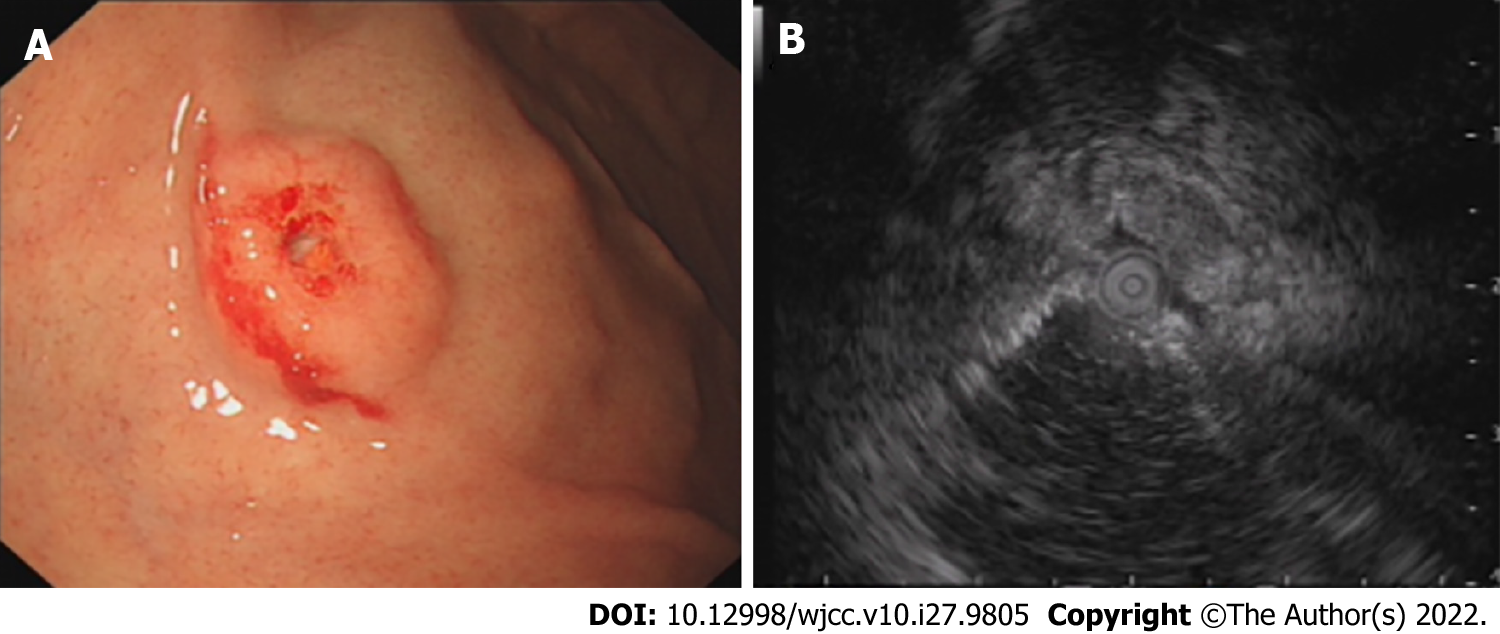
Figure 1 Endoscopic ultrasonography examination in the local hospital 1 year previously.
A: A solitary discoid-shaped submucosal tumor in the gastric fundus with central depression and surface mucosal congestion (IIa+IIc appearance, moderately irregular edges, presence of 2 mucosal folds converging on the lesion); B: The lesion originated from the deeper layers of the mucosa with partially discontinuous submucosa, medium and hypoechoic changes, and was 1.12 cm × 0.38 cm in size.
- Citation: Chen WG, Shan GD, Zhu HT, Chen LH, Xu GQ. Gastric metastasis presenting as submucosa tumors from renal cell carcinoma: A case report. World J Clin Cases 2022; 10(27): 9805-9813
- URL: https://www.wjgnet.com/2307-8960/full/v10/i27/9805.htm
- DOI: https://dx.doi.org/10.12998/wjcc.v10.i27.9805


