Copyright
©The Author(s) 2022.
World J Clin Cases. Sep 26, 2022; 10(27): 9776-9782
Published online Sep 26, 2022. doi: 10.12998/wjcc.v10.i27.9776
Published online Sep 26, 2022. doi: 10.12998/wjcc.v10.i27.9776
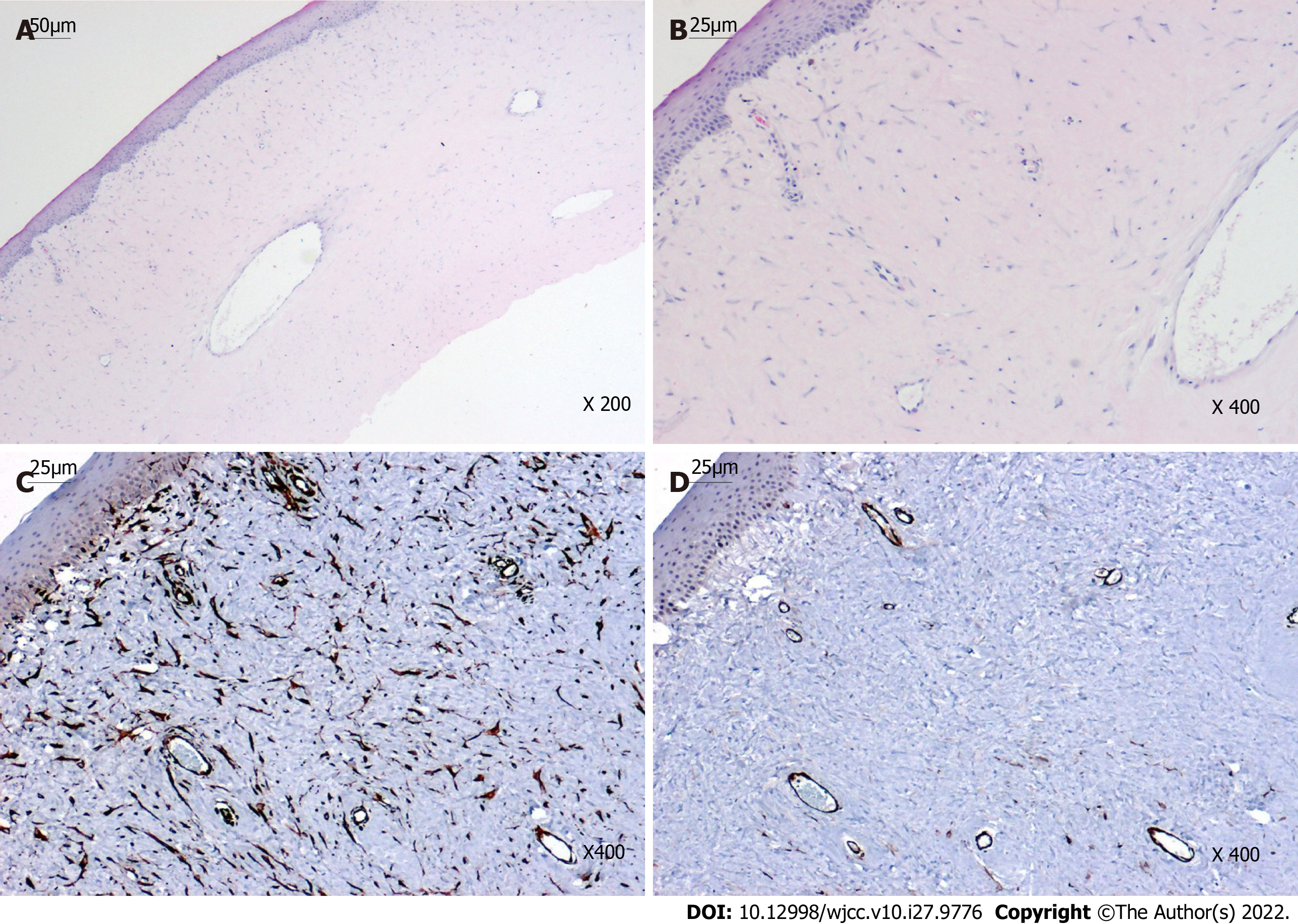
Figure 3 Histopathological and immunohistochemical results.
A-B: Hematoxylin and eosin staining showed the irregular surface of the swelling, non-keratinized epithelium without Bowman’s layer, and dense fibrous connective tissue with blood vessels beneath the epithelium; C: Vimentin staining was diffusely positive within the parenchyma of the mass; D: Smooth muscle actin staining was positive in the smooth muscle walls of the vasculature and myofibroblasts.
- Citation: Li S, Lei J, Wang YH, Xu XL, Yang K, Jie Y. Rare giant corneal keloid presenting 26 years after trauma: A case report . World J Clin Cases 2022; 10(27): 9776-9782
- URL: https://www.wjgnet.com/2307-8960/full/v10/i27/9776.htm
- DOI: https://dx.doi.org/10.12998/wjcc.v10.i27.9776


