Copyright
©The Author(s) 2022.
World J Clin Cases. Sep 26, 2022; 10(27): 9760-9767
Published online Sep 26, 2022. doi: 10.12998/wjcc.v10.i27.9760
Published online Sep 26, 2022. doi: 10.12998/wjcc.v10.i27.9760
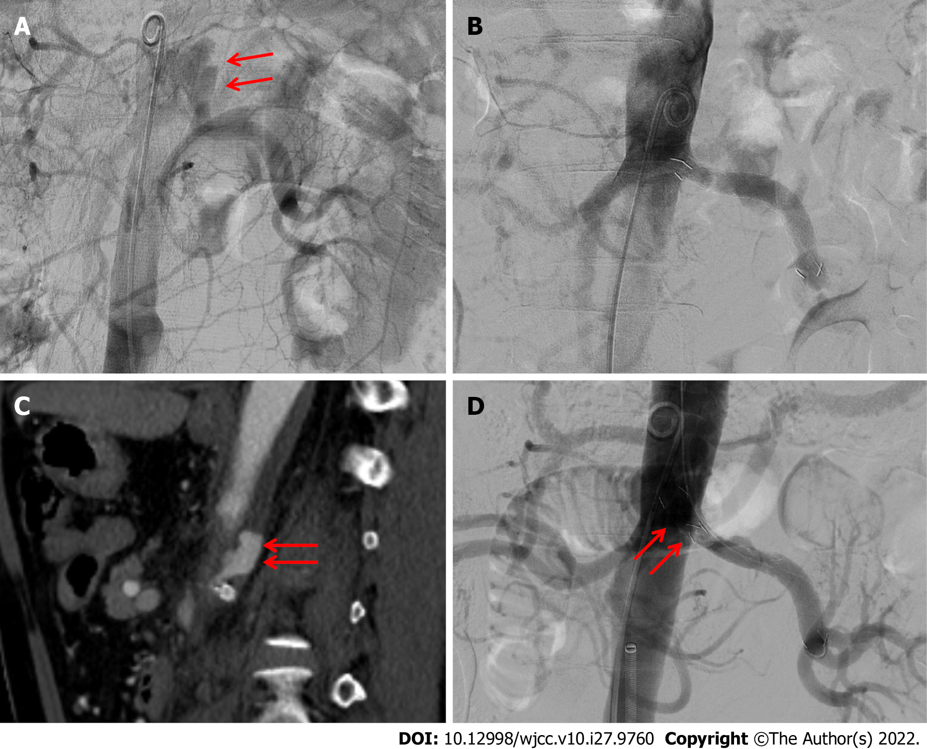
Figure 2 Endovascular procedure performed 15 d and 17 d after injury.
A: The pseudoaneurysm (red arrow) arising from the left proximal renal artery was detected using abdominal aortography 15 d after injury; B: An 8 mm × 55 mm covered stent was deployed along the left renal main artery, bridging the pseudoaneurysm and covering the parent artery, successfully excluding the pseudoaneurysm; exclusion was confirmed using final aortography; C: Follow-up CT scan two days after the first procedure reveals a decrease in the size of the pseudoaneurysm (red arrow) arising from the renal artery; D: Second endovascular procedure performed three days after the first treatment. The additional covered stent (red arrows) was placed to fully cover the orifice of the left renal artery to prevent 1A endoleak, successfully excluding the pseudoaneurysm, as confirmed using final aortography.
- Citation: Kim Y, Lee JY, Lee JS, Ye JB, Kim SH, Sul YH, Yoon SY, Choi JH, Choi H. Endovascular treatment of traumatic renal artery pseudoaneurysm with a Stanford type A intramural haematoma: A case report. World J Clin Cases 2022; 10(27): 9760-9767
- URL: https://www.wjgnet.com/2307-8960/full/v10/i27/9760.htm
- DOI: https://dx.doi.org/10.12998/wjcc.v10.i27.9760


