Copyright
©The Author(s) 2022.
World J Clin Cases. Sep 26, 2022; 10(27): 9750-9759
Published online Sep 26, 2022. doi: 10.12998/wjcc.v10.i27.9750
Published online Sep 26, 2022. doi: 10.12998/wjcc.v10.i27.9750
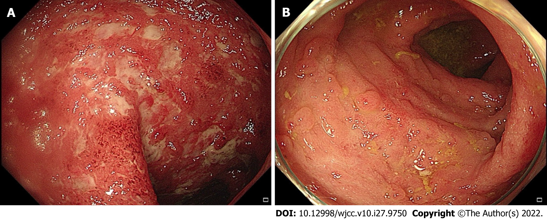
Figure 2 Initial endoscopic appearance of the rectosigmoid junction.
A: Extensive ulceration, diffuse erythema, and mucosal edema with loss of vascular markings; B: Endoscopy performed at 3-mo follow-up showing resolved ulceration and pseudopolyps.
- Citation: Wang YY, Shi W, Wang J, Li Y, Tian Z, Jiao Y. Myocarditis as an extraintestinal manifestation of ulcerative colitis: A case report and review of the literature. World J Clin Cases 2022; 10(27): 9750-9759
- URL: https://www.wjgnet.com/2307-8960/full/v10/i27/9750.htm
- DOI: https://dx.doi.org/10.12998/wjcc.v10.i27.9750


